| |

| 
| |
Spinal Anatomy 101
This section was compiled by Frank M. Painter, D.C.
Send all comments or additions to:
Frankp@chiro.org
|
 |
 |
| |
| |
|
| |
|
|
|
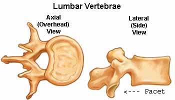
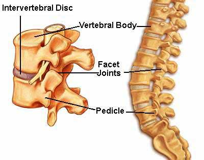
This is a representation of a lumbar vertebra. On the right-side of both sketches is the vertebral body. On the left-side of both views is the ring of bone (called the lamina) which houses the spinal cord.
The lamina attaches to the vertebral body by the notched pedicle.
The rounded space (or notch) formed just above and below the pedicle creates the intervertebral foramina (IVF).
Directly behind the pedicle lies the articular pillar.
At the top and bottom of each articular pillar are found the facet joints (zygapophyseal or “Z joints”) which permit the functional motions of the vertebra, including flexion, extension, rotation, and lateral bending.
|
|
|
|
| |
| |
|
| |
|
|
|
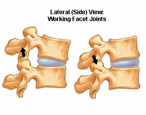
The “Spinal Motion Unit”
Two vertebra, and the tissues that connect them, comprise the smallest working unit of the spine. This unit is referred to as the “spinal motion unit”. The motion unit shown on the left demonstrates normal flexion and extension.
You will also notice that the only motion that takes place at the vertebral disc (IVD) is compression at the front of the disc (during flexion), and at the back of the disc (during extension).
There should be no forward or backward motion of the vertebral bodies (also known as translation) when the integrity of the IVD remains intact.
See the hole directly behind the blue disc? This is the intervertebral foramina (IVF) and this is where the spinal nerve roots pass from the spinal cord, out to the body. When the vertebra flexes, the facets slide upwards and forwards, and the size of IVF increases. In extension, the size of the IVF decreases.
|
|
|
|
| |
| |
|
| |
|
|
|
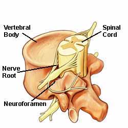
Nerve Root Impingement
Nerve roots, as they exit the foramina (or IVF), pass between the disc and the facet complex (see upper picture). While in the IVF, they face the dual challenge known as being “pickle in the middle”.
The facet complex is located directly behind the foramina (IVF). If there is inflammation at the facets, the swelling of the joint capsule may encroach into the foramina, impinge upon the delicate nerve root fibers, and cause nerve root irritation (also known as “radiculopathy”).
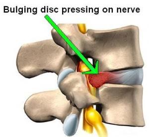
The disc is located directly in front of the IVF. The picture on the bottom demonstrates nerve root impingement being caused by a herniated disc. This may also cause radicular symptoms.
The typical symptoms of radiculopathy include pain, tingling or numbness (“paresthesia”), and in the most extreme cases, muscle weakness along the distribution of that nerve root.
|
|
|
|
| |
| |
|
| |
|
|
This sketch shows the nerve root exiting the IVF, and perhaps makes it easier to visualize how inflammation, either at the facet joints or the disc, can encroach upon the delicate nerve roots.
|

Return to SUBLUXATION ANATOMY
Since 5-16-2007
Updated 3-29-2025
|
|
|
|
|