
|
|
Chapter 10
Introduction to Chiropractic Physiologic Therapeutics
From R. C. Schafer, DC, PhD, FICC's best-selling book:
“Basic Chiropractic Procedural Manual”
The following materials are provided as a service to our profession. There is no charge for individuals to copy and file these materials. However, they cannot be sold or used in any group or commercial venture without written permission from ACAPress.
All of Dr. Schafer's books are now available on CDs, with all proceeds being donated
to support chiropractic research. Please review the complete list of available books.
Introduction

Definitions and Rationale

Synopsis of Physiotherapeutic Procedures Relative to Pathophysiology
  Massage
    Physiologic Effects
    Indications
    Contraindications
  General Rules of Application
  Massage Media
  Common Massage Techniques
    General Effleurage
    Fulling Effleurage
    Knuckling Effleurage
    Shingling Effleurage
    Bilateral Tree Effleurage
    Petrissage (Compression)
    Tapotement (Percussion)
    Vibration
  Reflex Effects of Massage
  Mechanical Effects of Massage

Therapeutic Traction
  Physiologic Effects of Continuous Moderate Traction
  Physiologic Effects of Intermittent or Alternating Traction
  Contraindications of Continuous and Intermittent Traction
    Further Contraindications to Intermittent Traction
  General Rules of Application

Mechanical Supports (Braces, Casts, Shoelifts, etc)
  Physiologic Effects
  Contraindications
  Basic Rules of Application
  General Considerations in the Use of Braces or Jackets
  Spinal Braces
  Molded Jackets
  Applications of Rigid Supports for the Back
  Belts
  Aspirated Air Belts
  Corsets
  Extremity Supports
  Considerations in Chronic Disorders
    Regional Mobilization
    General Objectives in Joint Support
    Muscle Re-education

Cryotherapy
  Physiologic Effects of Cold
    Local Consequences
    General Consequences
  Indications for Local Cooling
  Contraindications to Local Cooling
  General Rules for Applying Cold
  Plastic Bag Pack
  Vapor Coolant Sprays
  Ice Massage
  Extremity Immersion Cold Bath
  Cryokinetics
  Combined Local Cooling and Remote Heating
  Cold Wet Packs
  Contrast Baths

Infrared and/or Therapeutic Heat
  Physiologic Effects of Local Heat
    Local Consequences
    General Consequences
  Contraindications to Local Heat
  General Rules of Application for Local Heat
  Infrared or Radiant Heat Radiation
  Hydrocollator Packs
  Hot Wet Packs

Shortwave Diathermy
  Major Indications
  Precautions
  Physiologic Effects
  Contraindications
  General Rules of Application

Microwave Diathermy
  Indications
  Contraindications
  Application

Ultrasonic Diathermy
  Physiologic Effects
  Contraindications
  Intensity
  Time
  Application Considerations
    The Coupling Medium
    Oil Coupling Agent
    Underwater Technique
    Phonophoresis

Sinusoidal Current
  Physiologic Effects
  Contraindications
  General Rules of Application

Galvanic Current
  Dosage
  Physiologic Effects
  Polar Effects
    Positive Pole
    Negative Pole
  Contraindications
  General Rules of Application With or Without Iontophoresis
    First Steps
    Pad Covers
    Technique
  Current Regulation
    Interrupted Galvanic Current
    Faradic Current
    Electric Stimulation
      Denervated Muscle
      Innervated Muscle
      General Rules of Application

Iontophoresis
  Substances Commonly Used
    Positive Pole
    Negative Pole
  Application Considerations
  Therapeutic Uses
  General Rules in Application

Ultraviolet Therapy
  Minimal Erythema Dose
  Physiologic Effects
    Local Erythema Dose
    Pigmentation and Metabolic Effects
    Bactericidal Effects
    Counterirritation Effects
  General Rules of Application
  General Indications
  Contraindications

Therapeutic Exercise
  Background
  The Need for Mobility Maintenance
  The Effect of Restricted Mobility in Maintaining Equilibrium
  The Hip, Knee, and Ankle During Gait
  Maintaining and Increasing Ranges of Motion by Stretching
  Neuromuscular Re-education
  The Development of Strength, Endurance, and Power

Electromyography
  The Neurophysiology Involved
  Instrumentation
  Areas of Application
  Traditional Electrodiagnostic Techniques vs Electromyography

Meridian Therapy
  Physiologic Effects of Acupuncture
  Complications of and Contraindications to Acupuncture
  The Rationale
  The Energy System
  Basic Concepts of Application
    Terminology
    Obstacles and Stimulants to Energy Flow
    Analysis and Redirection of Energy
    Major vs Minor Roles in Therapy
Concluding Remarks
Chapter 10: Introduction to Chiropractic Physiologic Therapeutics
The use of physiotherapy and physical therapy to enhance the effects of the chiropractic adjustment in treatment can be significant in many cases. Superficial heat, diathermy, cold, microwaves, ultrasound, ultraviolet rays, galvanic and sinusoidal currents, traction, hydrotherapy, or therapeutic massage and exercise are among the therapies that may benefit the patient when properly applied. These procedures may help to reduce stiffness in joints, relieve tension, relax muscle spasm, and offer many other physiologic benefits.
Special precautions, however, must be observed when treating patients of advanced age. Special consideration must also be given to indications and contraindications, patient sensitivity, intensity, and duration of treatment.
Special caution must be used with patients that have heart and blood pressure problems, renal failure, diminished sensation or circulation, or an inability to tolerate heat or cold. For example, patients with Raynaud's disease do not tolerate cold. Patients with other circulatory problems do not tolerate thermotherapy because they have less ability to dissipate the heat. Patients with a distinct loss of sensation will not realize if an area is being overheated or even being burned.
A patient's tolerance cannot be the only guide to intensities and duration of treatment. Frequent checking, both visually for redness and by palpation to determine over heating, must be done during the treatment period. Reasonable examination, monitoring, and care by the doctor can avoid problems in most instances.
INTRODUCTIONPhysiotherapy techniques are frequently used preparatory to the chiropractic adjustment to improve function, relieve spasm, minimize pain, and enhance circulation and drainage. They are often used before primary care to relax the patient and condition tissues, and posttherapy to relive pain and prevent deformities resulting from trauma or disease and to maintain what has been gained in treatment. There are also times when it may be considered primary therapy. Rehabilitation objectives are shown in Table 10.1.
While this chapter has not made an attempt to describe the physics involved for each modality, both practitioner and therapist should be well acquainted with underlying fundamentals to prescribe appropriate procedure, intensity, duration, and technique, as well as effectively analyze methodology and evaluate treatment.
The roles several procedures play in chiropractic physiologic therapeutics are described such as for massage, traction, mechanical supports, colonic irrigation, dietetics, cryotherapy, heat, infrared radiation, shortwave diathermy, microwave diathermy, ultrasonic diathermy, sinusoidal currents, galvanic current, iontophoresis, ultraviolet therapy, therapeutic exercises, and acupuncture.
As techniques in application vary extensively with the equipment used, it is important that both practitioner and therapist be fully acquainted with the equipment manual furnished by the manufacturer. In purchasing equipment, the physician must assure that the equipment meets all government and professional standards.
Table 10.1. Basic Functional Goals in Rehabilitative TherapyFunction Dysfunction Primary Therapy Strength Weakness Exercises against resistance Physiologic Spasticity Thermotherapy, ultrasound, autosugges- elasticity tion, biofeedback therapy, postural correction, relaxing exercises Spasm Pain relief, "gate" blockage tech- niques, relaxing exercises, heat Tension Relaxing exercises, hydrotherapy, bio- feedback, hypnotherapy, psycho- therapy Physical Contracture Stretching exercises, joint mobiliza- elasticity tion, ultrasound, thermotherapy Coordination Incoordination Strengthening and relaxation exer- cises, coordination training and practice.
DEFINITIONS AND RATIONALEChiropractic physiotherapy is the therapeutic application of forces and substances that induce a physiologic response and that uses and/or allows the body's natural processes to return to a more normal state of health. It makes use of the therapeutic effects of:
Soft tissue manipulation and massage. The latter may include stroking, compression, percussion, vibration, mild passive joint motion, nerve stimulation, and several forms of reflex and trigger-point therapy.
Mechanotherapy, including active and passive exercise; traction, either intermittent or sustained; and structural braces, shoe lifts, casts, or other supports.
Light, heat, cold, air, and water are natural forces that are the prerogatives of all people to be free to apply.
Hydrotherapy, in its traditional forms.
Electrotherapy, when therapeutic procedures are in keeping with a justified physiologic intent and reaction.
Nutritional planning, dietetics, and special food or nutritional supplementation. These have been described in Chapter 9.
The rational use of the forces and substances shown in Table 10.2 first requires an understanding of their mechanism of action and their predictable effects on pathophysiologic processes and on tissues with impaired function and tolerance. Second, an understanding of the abnormal or diseased state must be determined; ie, the quantity and quality of the abnormal processes involved at the time of therapy are the primary criteria for proper application.
In contrast, a diagnostic entity may at various times in its course entail several of abnormal physiologic reactions and processes; therefore, treatment must vary with the abnormal process and be adapted to it rather than statically limited by a specific unalterabIe concept of a disease entity. Disease is not a thing, but a process --a normal process out of time or phase, and the primary intent of chiropractic physiotherapeutics is to help the body adapt to and/or normalize the detrimental processes of a diseased state.
Table 10.2. Brief Resume of Common Physical Agents and Their Effects
Physical Agent                 Primary Effect      Secondary Effects
Hot water, hot air, radiant    Thermal             Hyperemia, sedation of sensory
heaters, incandescent lamps,                       or motor irritation, attenua-
diathermy, microwaves                              tion of microorganisms

Cryotherapy (vapocoolants,     Hypothermal         Sedation, decongestion, ischemia
ice)

Ultraviolet (sun, heated       Photochemical       Erythemia, pigmentation, acti-
metals, carbon arc, mercury                        vation of ergosterol
vapor arc)

Ultrasound                     Mechanical, ther-   Cellular massage, heat, sedation
                               mal, chemical

Low voltage galvanic           Electrochemical     Polar, vasomotor
currents

Low-frequency interrupted      Electrokinetic      Muscle stimulation, increase
current, sinusoidal current,                       of venous and lymph flow, re-
other alternating currents                         flex stimulation

Vibration, massage, traction   Kinetic             Muscle stimulation, increase
(intermittent), therapeutic                        of venous and lymph flow,
exercise                                           tissue stretch, reflex stimu-
                                                   lation
Any physiotherapeutic process must parallel the concomitant pathogenesis
and be directed at stopping and/or reversing the process as it currently
exists. For these reasons, it is advantageous to review briefly the
pathophysiologic processes from the initial trauma to the extreme of fibrosis.
Whether a tissue becomes primarily injured through direct overt trauma or
microtrauma, or is undergoing a change as a secondary reaction to a pathologic
process initiated elsewhere, the following stages usually occur: hyperemia,
passive congestion, consolidation of the protein exudate, formation of a
fibrinous coagulate, organization through fibroblastic activity, fibrosis,
ischemia, and, therefore, possibly more fibrosis and/or atrophy. See Table
10.3.
These processes often exist in different ratios within the same tissue at
the same time. However, one usually dominates; and treatment should be
directed primarily for it. Changes or modifications in therapy are often
necessary as the dominant feature alters.
The presence of a coexisting neuropathy must also be realized. The area of
therapy should be considered as not only the local tissue but also the
neuromere or spinal segment directly involved. If one relates this process to
the physiotherapeutic measures and their effects, it is understandable how
appropriate these procedures may be.
| I. Stage of Hyperemia or Active Congestion |
Ice packs: vasoconstrictive effects.
Galvanism: vasoconstrictive, hardening of tissues effects.
Pulsed ultrasound: dispersing effects; increased membrane permeability effects.
Rest, with possible support: prevents irritation and further injury.
| II. Stage of Passive Congestion |
Alternating hot and cold applications, preferably in a 3:1 ratio every few hours: revulsive effects.
Light massage, particularly effleurage: revulsive effects.
Passive manipulation: effects of revulsion, maintenance of muscle tone, freeing of coagulate and possibly early adhesions.
Mild range of motion exercise: effects same as 3.
Alternating current stimulation, of a surging nature: effects same as 3.
Ultrasound: increase in gaseous exchange, dispersion of fluids, liquefaction of gels, and increased membrane permeability effects.
| III. Stage of Consolidation and/or Formation of Fibrinous Coagulant |
Local moderate heat, preferably of a moist nature: mild vasodilation, increased membrane permeability effect.
Moderate active exercise: revulsive effects, freeing of coagulant and early adhesions, maintenance of tone, and ligamentous and muscular integrity effects.
Motorized alternating traction: effects same as 2.
Moderate range of motion manipulation: effects same as 2.
Ultrasound: hyperemia, liquefaction of gels, dispersion of gases and fluids, increased membrane permeability, and tissue-softening effects.
Sinusoidal current, surging or pulsating: effects same as 2.
| IV. Stage of Fibroblastic Activity and Fibrosis |
Deep heat, prolonged (eg, diathermy): prolonged vasodilation, increased membrane permeability, increased chemical activity effects.
Deep massage (eg, petrissage or other soft-tissue manipulation: tends to break down fibrotic tissue and create more elasticity.
Vigorous active exercise, preferably with slight traction or at least without weight bearing: maintains muscle and ligamentous integrity, stretches fibrotic tissues, breaks adhesions, and creates greater elasticity.
Motorized alternating traction: effects same as 3.
Negative galvanism, particularly with an antisclerotic (eg, potassium iodine): vasodilation, softening, liquefaction, and antisclerotic activity effects.
Ultrasound: effects causing softening of tissues as previously described.
MASSAGE
Therapeutic massage provided by a professional should not be confused with
that given by an unlicensed practitioner. Scientifically applied massage acts
mechanically on the tissues that are directly and reflexly contacted on deeper
or possibly remote visceral structures. When applied to stretch adhesions and
contractures or to relocate prolapsed organs, the term "bloodless surgery" is
sometimes used.
Physiologic Effects
Massage increases the flow of blood and lymph; increases pulse beat;
raises blood pressure when moderately forceful; reduces edema, congestion, and
exudates; reduces joint effusions and coagulates; stretches and/or breaks
tissue and interseptal adhesions; increases urinary production; increases
respiration when moderately forceful; sedates motor and sensory nerve
activity; and removes lactic acid from musculature and relieves fatigue. When
applied to the abdomen, it increases peristalsis and the elimination of
intestinal residues and debris.
Indications
Massage is especially useful in conditions in which relief of pain,
reduction of swelling, or mobilization of contracted tissues is desired. It
is almost always indicated in posttraumatic swelling and induration such as
seen in strains, sprains, subluxations, bruises, and tendon and nerve
injuries. It is often used in such disorders as arthritis, periarthritis,
bursitis, neuralgia, fibrositis, spastic low-back pain, paralytic conditions,
and psychoneurosis.
Massage does not increase muscle strength and is not a substitute for
exercise. Strength develops in muscles contracting actively, preferably
against resistance.
Contraindications
Typical contraindications include skin infections and ulceration, local
acute inflammatory processes, adiposis dolorosa and other lipodystrophies,
abrasions, cuts, acute deep or visceral inflammations, phlebitis, thrombosis,
and other vascular diseases where there is a tendency to hemorrhage such as
with varicosities, peptic ulcers, a menstruating uterus, acute osteomyelitis,
tubercular joints, any infectious bone or joint disease, or malignant
neoplasias.
General Rules of Application
Adequate lubricant should be used to avoid irritating the skin. Too
vigorous or traumatic forces, or too lengthy a treatment time must be avoided.
Blood vessels, inflamed nerves, or other sensitive structures or areas should
be avoided during deep stroking or effleurage. Regional massage should be
applied centripetally toward the heart.
Prior to treatment, inform the patient what you are going to do, how you
are going to do it, and why it is necessary. This avoids patient anxiety and
enhances cooperation.
Massage Media
Lotions, creams, oils, and other lubricants are primarily used to reduce
friction and soften skin or scar tissue. Powders such as talcum make kneading
strokes easier, but avoid powders on the faces of patients with respiratory
problems.
Before applying the medium, clean the patient's skin at the application
site, inform the patient about the medium, and assure the patient is not
sensitive to the medium. Following therapy, remove the medium with alcohol if
indicated but never pour alcohol directly on the skin. Inspect the area after
cleaning and record findings.
Common Massage Techniques
General Effleurage
Essentially, effleurage means stroking the skin with light or heavy
pressure. On large areas, the hands are relaxed, the fingers are together, and
the palms serve as the primary contact point. On small areas, the thumbpads
and/or fingerpads are used. Movements are long centripetal strokes where
skin contact is maintained at the end of each stroke when returning to the
starting position. Mild effleurage is often used as a preliminary to, between,
or to conclude other massage techniques.
Fulling Effleurage
Using the palms and fingers, one hand pushes with this technique while the
other hand pulls. This procedure is frequently useful in the lower back and on
large muscle areas.
In low-back musculoskeletal conditions, place one hand on the far side
from the spine, pointing fingers laterally. Place the other hand with fingers
pointing medially on the near side of the spine. The far hand is pulled toward
you as the near hand is pushed away from you while avoiding pressure on the
spinous processes as the hands meet and cross to the opposite side.
Knuckling Effleurage
Very deep effleurage is accomplished by making a fist and placing the
dorsal surface of the first phalanges on the part to be massaged and then
forcefully extending the wrist in a flicking motion. Movements proceed in a
centripetal direction.
Shingling Effleurage
This type of effleurage is when one stroke is made on top of the previous
stroke, like over-lapping shingles on a roof. One hand is placed on the part
and moved toward you. At the completion of this movement, the other hand is
placed where the first hand started and moved toward you. As one hand
completes the stroke, the other hand begins a stroke. Direction is up the part
(cephalad) such as on a limb or the spine.
Bilateral Tree Effleurage
This is a common spinal technique where the hands are placed on the lower
back; the fingers are together and point medially with the palms next to but
not on the spinous processes. The hands are removed after each lateral stroke,
and the direction of the effleurage progresses up the spine.
Petrissage (Compression)
This is a muscle kneading technique where little or no lubricant is used.
The hand(s), rather than sliding over the skin, contacts the muscle and rolls
and compresses it against bone or another structure. Tissues are sometimes
alternately kneaded between the fingers of one hand and the thumb of the other
hand.
Tapotement (Percussion)
This is a series of rapid percussion strokes by:
Tapping with the volar surface of the fingers extended, alternating hands in rapid succession.
Slapping with palm and fingers, alternating hands in rapid succession.
Hacking in rapid succession with palms facing each other, fingers relaxed, and using the ulnar borders of the hands during contact.
Thumping, similar to hacking but with hand made into a fist.
Cupping, slapping with the palms in a cupped position, fingers semiflexed.
Pincemont, a gentle pinching of tissues between the thumb and index finger in rapid succession.
Vibration
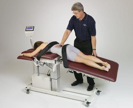
Traction tables can be designed to apply continuous traction and/or
intermittent traction combined with heat, vibration, and/or massage. See
Figure 10.1.
Immobilizes and "splints" injured or overstressed musculoskeletal
tissues.
Relieves spastic muscles by placing them in "physiologic rest."
Relieves compression effects on articular tissues due to spasm or other
pressure-inducing factors.
Reduces circumference of intervertebral disc. Therapy helps to restore
normal positioning.
Relieves compression effects of foramenn distortion and/or narrowing.
As a by-product of the previous features, it helps to dissipate
congestion, stasis, and edema of associated tissues.
Stimulates proprioceptive reflexes.
Stretches fibrotic tissues and adhesions. Increases vascular and lymphatic flow; thus reducing stasis, edema, and
coagulates in chronic congestion.
Helps "tone" muscles, thereby reducing fatigue and restoring tissue
elasticity and resiliency.
Stretches and helps free periarticular and articular adhesions and
fibrotic infiltrations.
Encourages expansion and contraction of disc tissues; thereby improving
the nutritional media.
Stimulates proprioceptive reflexes.
Local osseous infections such as osteomyelitis, tuberculosis, etc.
Osteoporosis and/or osteomalacia.
Osseous neoplasias.
Severe cardiovascular and/or hypertensive disease.
Localized vascular disease and/or with a tendency for hemorrhage in the
area.
Advanced cachexia.
Pregnancy (in areas that might adversely affect the gravid uterus).
Diseases of the spinal cord.
Inflammatory and/or rheumatoid arthritis.
Severe spasms.
Acute inflammations of musculoskeletal tissues such as myofascitis,
bursitis, tendinitis, etc.
Acute intervertebral disc syndrome.
General Rules of Application
The hand(s) is placed against the skin, and a tremulous movement is
applied.
Reflex Effects of Massage
Cutaneous reflexes are produced in the skin by stimulating peripheral
receptors that initiate impulses that are conducted via sensory neurons to
the spinal cord and ascending tracts to the brain. The result is sensations of
relaxation or pleasure. Peripherally, these impulses cause relaxation of
muscles and dilatation or constriction of arterioles. Sedation occurs when the
massage is given in a monotonously repetitive manner without sharp variations
in pressure or irritating changes in application. Relaxation of musculature
and reduction of mental tension are other common effects.
Mechanical Effects of Massage
The mechanical effects of massage result from measures assisting
circulation and drainage of blood and lymph because the movements are given
with the greatest force in the centripetal direction along with measures
producing intramuscular motion. The stretching of adhesions between muscle
fibers and the mobilization of fluid accumulations also are mechanical
effects.
THERAPEUTIC TRACTION
Physiologic Effects of Continuous Moderate Traction:

Physiologic Effects of Intermittent or Alternating Traction:
Contraindications of Continuous and Intermittent Traction:
Further Contraindications to Intermittent Traction:
 |
Before application, explain to the patient the procedure that will be
used, what the patient should feel, and why this particular procedure is used.
Periodically, recheck the angle of pull and patient comfort. See Figure 10.2.
Place the patient in a position that will best influence the area of
treatment. Pads, pillows, and similar devices are often helpful. Place
attachment halters, straps, or other traction connections firmly but with
ample padding to the patient's skin, soft tissues, or osseous structures. See Figure 10.3.
Physiologic Effects
Weight should be increased and/or decreased slowly to avoid abrupt
reactions. The amount of weight should be measured by a superimposed scale and
adjusted to the patient's condition, age, size, strength, health, etc.
Note: Generally, 5% of the patient's body weight will allow a separating
effect in the cervical spine, and 25% of the body weight will allow a
separating effect in the lumbar spine.
The duration of therapy must be adapted to the condition treated; however,
undertreatment is better than excessive therapy. Time can always be increased
after a trial period.
See that the patient is well attended and monitored. Cease therapy if
adverse symptoms or signs develop.
MECHANICAL SUPPORTS (Braces, Casts, Shoelifts, etc)
 |
Mechanical supports hold structures in a sustained position and thus
promote healing. See Figure 10.4. They relieve weight-bearing or motion stress
on joints and osseous structures. They relieve muscle, tendon, and ligament
stress from postural and/or motion efforts.
Some supports such as shoelifts allow stretching and/or contracting of
musculoskeletal tissues and thus encourage structural alterations. In
anomalous and/or acquired malformations, shoelifts and other supports may
provide for a structural deficiency of formation.
Contraindications
Contradictions arise where immobilization may promote the organization of
inflammatory coagulates and consequent adhesions and/or fibrotic infiltration
--depending on time and nature of the condition. Immobilization can also
encourage muscular atonicity weakness and/or atrophy --depending on time and
nature of the condition.
Immobilization or a sustained position may promote unsatisfactory
stretching and/or contractual changes --depending on time and nature of the
condition. Immobilization can also cause vascular stasis, congestion, or
ischemia --depending on time and nature of the condition.
Basic Rules of Application
 |
Be certain of patient comfort yet firm fitting of the device. See Figure
10.5. Hold the part in proper position, usually that of "physiologic rest."
Avoid friction or other irritation of the skin; avoid pressure on blood
vessels, nerves, or other vital structures; avoid pressure that may cause
impairment to venous or lymphatic drainage; initially allow for swelling; and
avoid stress on ligaments, muscles, tendons, or other tissues.
Watch for adverse symptoms such as pain, pallor, pulselessness,
paresthesia, or swelling. Periodically check and re-evaluate support and its
function. When possible, use periodic mobility, passive or active exercises,
or any other proper therapy to counteract the deleterious side effects of
immobilization. Much of this can be minimized by passive exercise (manual,
mechanical, electrical) of adjacent joints.
General Considerations in the Use of Braces or Jackets
Braces or jackets are applied to the body for support or immobilization of
a specific part or region to correct or prevent deformity and to assist or
restore function. Common indications for braces are local pain, weakness,
degeneration, or paralysis.
In prescription, it should be kept in mind that one of the basic
principles of design is to provide adequate surface area for the distribution
of forces involved. Accurate contouring helps.
If immobilization of a joint is desired, the brace will require long lever
arms with maximum pressure distribution to give comfort and be effective.
Proper design should consider simplicity, durability, minimal weight,
necessary strength, and be as inconspicuous as possible for cosmetic reasons.
Prescription for a support or device should be specific, recorded in the
patient's record, and should include the part(s) to be braced, type of brace,
material, type of joints, and special considerations, After delivery, the
brace should be checked to assure that the prescription has been filled
properly and that it fits the patient adequately.
Progressive therapeutic exercises, both passive and then active, should be
used during and following the use of braces.
Spinal Braces
Spinal braces have been used widely for hundreds of years for both
corrective and supportive goals such as relief of pain, prevention and
correction of deformity, protection against further injury, and as an aid in
supporting weakened muscles and ligaments.
Any involved spinal biomechanics should be understood before spinal braces
are considered. Remember that the spinal column acts as a flexible elastic rod
for maintaining the upright position where intrinsic stability is provided by:
(1) the discs,
(2) muscular support, and
(3) ligament straps. The compression,
shearing, twisting, and bending forces withstood in daily living are the
result of these combined intrinsic factors and extrinsic loading.
Because of the intervertebral discs, the spine as a whole is quite
flexible, elastic, and plastic. When the anulus fibrosis is intact and the
nucleus has not been displaced, the elastic limits by compressive forces
cannot be exceeded without vertebral fracture. The end-plate will usually
fracture before the vertebral body unless advanced bone disease exists that
predisposes bone failure (eg, osteoporosis, cystic cavitations, etc).
The spine can be considered a segmental elastic column supported by the
paraspinal muscles attached to the sides and located within the abdominal and
thoracic cavities. Trunk muscles convert these cavities into rigid-walled
cylinders capable of transmitting forces developed in loading the spine and
thus relieve some pressure on the spine itself. For instance, the
greater the weight lifted, the greater the activity of the trunk, chest, and
abdominal muscles.
Without trunk support and cavity pressures, forces on the lumbosacral disc
would be 30% greater and those on the lower thoracic spine would be about 50%
greater. A tight corset about the abdomen leads to less abdominal and thoracic
muscular activity when lifting, indicating that the effect of these muscles
can be at least partially replaced by external support.
Both gait and energy expenditure are influenced by spinal appliances. A
patient with an appliance restricting axial spinal rotation walks slower with
shorter steps in order to maintain a comfortable pace, maintain balance, and
conserve energy.
In the use of any spinal appliance, spinal motion is increased in the
segments adjacent to the end of the appliance. This may be purposeful in
design; if not, resulting discomfort may require extension of the brace well
above and below the involved area to relieve discomfort.
Except for a hyperextension brace, almost all spinal appliances depend
partially on compression to achieve their goal. In spinal support, it is
generally assumed that relief from back pain is the result primarily of
abdominal compression and secondarily from the decrease in lumbar lordosis.
Molded Jackets
Jackets made of plaster of paris, leather, or plastic are molded to fit
the contour of the body and assist in distributing even pressure. They are
often useful in providing spinal support for debilitated patients in
degenerative disorders such as osteoporosis, chronic poliomyelitis, and severe
spinal deformities and congenital anomalies, and to restrict movement after
acute strain or sprain. See Figure 10.6.
Applications of Rigid Supports for the Back
Commonly used in cases of back pain, a rigid spinal brace often provides
an efficient method of obtaining abdominal compression with an anterior force
and distributing the counterforce over a wide area when properly fitted with
the rigid uprights contoured to follow the lumbar curve.
Manufactured in several styles, the rigid back support essentially
consists of a pelvic band at a level between the greater trochanter and the iliac crest and a thoracic band at the level of T8. These bands connect by two
lateral and posterior uprights and usually with a corset front.
The typical dorsolumbar brace is a long brace consisting of a wide-fitted
pelvic band and two long posterior paraspinal uprights extending to the
shoulders. The uprights are connected in the midthoracic area by a short
transverse bar, with straps from the uprights passing over the shoulders and
under the axillae to the transverse bar. A full-length abdominal apron is
often attached.
If the goal is to immobilize, a rigid support should be used that is
designed and fit to accomplish this purpose (eg, supports using plastics,
metal, and/or leather). If the goal is to allow slight mobility after
inflammation has subsided, then a support allowing some motion can be used
(eg, fabrics or materials offering some elasticity, flexibility, and/or
plasticity such as foam collars or elastic bandages).
Belts
While corsets are commonly prescribed for women, belts are more common for
men. The substitution of the common elastic belt is strictly to satisfy the
male ego for the corset is by far a more efficient support.
In sacroiliac inflammatory or hypermobile disorders, a trochanteric belt,
usually from 2 to 3 inches wide, is worn around the pelvis between the
trochanters and iliac crests. However, this type of appliance may offer less
than ideal support because of a too narrow width.
Sacroiliac and lumbosacral belts are similar in design and function,
differing essentially only in width. The sacroiliac belt is usually from 4 to
6 inches wide, while the lumbosacral belt is usually from 8 to 16 inches wide.
Both sacroiliac and lumbosacral belts are often useful in back pain resulting
from disc disorders; however, they fail to provide sufficient immobilization
in extremely severe conditions unless they are made of inflexible and
inelastic material and incorporate metal stays.
Aspirated Air Belts
A relatively new product to protect and support the lower back, especially
as an aid during rehabilitation following low-back trauma (sprain and/or
strain) is the inflatable "air belt." An air belt is a uniquely designed
light-weight yet strong lumbar support that can be worn either inside or
outside of clothing. It incorporates many advantages of a heavy inflexible
corset yet is lightweight and "is automatically custom-fit" to conform to the
patient's unique body shape. Unlike rigid corsets, motions of normal daily
living are not restricted when it is worn and the patient has immediately
control of the pressure.
The examples shown in Figures 10.7 and 10.8 are designed so interconnected
chambers in the belt allow conformity to the patient's spine. These supports
have a Camrelle inner liner for moisture absorption and a durable outer shell
to provide rigidity. As the chambers are aspirated with air by a "tuck away"
palm grip bulb, the chambers expand and apply forward pressure over the muscles of the lower back. This relaxes stressed spinal muscles and ligaments
and encourages proper biomechanical balance and segmental alignment.
Corsets
Corsets are encircling garments having vertical and/or horizontal
metal stays. They generally encase the region between the pubis and lower
ribs. The higher the back of the corset, the greater spinal support.
Corsets are often prescribed to assist weakened muscles or to limit spinal
motion. In general, a corset is worn only during active hours, but in cases of
severe low back pain, it may give relief if worn both day and night. As
always, prolonged constant support produces weakened muscles and ligaments and
encourages fibrosis.
 |
Extremity Supports
A large variety of elastic and hinged appliances are available to support
and/or immobilize an injured shoulder, elbow, wrist, knee, or ankle. Taping
is another common technique used to achieve the same objectives. Examples are
shown in Figure 10.9 and Figure 10.10.
Considerations in Chronic Disorders
In addition to chiropractic articular adjustments, procedures involving
regional mobilization, articular support, and muscle re-education are often
highly beneficial. Nevertheless, the use of supportive appliances is always a
matter of weighed clinical judgment. When used wisely with a thorough
knowledge of biomechanics, application may be highly beneficial as an adjunct
to corrective adjustments in many acute and chronic disorders.
Note: Immobile support without the advantage of prior mobilizing
adjustments and not used in conjunction with neuromuscular re-education rarely
achieves optimal goals.
Regional Mobilization
The goal of regional mobilization by external appliances is to regain or
maintain mobility of the affected region by enhancing the correction of
pertinent articular fixations and obtaining symmetric mobility for successful
muscle reeducation and support by bracing.
Muscle stretching is often helpful, but it is essentially useless and
possibly contraindicated in tightening tissues on the convexity of a
scoliosis. Manual or mechanical muscle stretching is effective and should be
specific for the concavity of the low thoracic, thoracolumbar, and upper
lumbar regions. Unfortunately, it is rarely beneficial in stubborn fixations
in the upper thoracic region or at lumbosacral angle. It is most effective in
all regions during the formative stages but rarely helpful after bony changes
have occurred. Nevertheless, any degree of restricted articular mobility that
can be restored will be of benefit to the patient.
Recommended isotonic or isometric exercises should be done gently (to
tolerance) and repeated several times a day. Stretching by a properly designed back brace or corset has limited value in correcting lumbosacral and high
thoracic curves, but intermittent spinal traction may be helpful.
General Objectives in Joint Support
The aims of corsets, braces, adjustable frames, and casts are fourfold:
(1) to support weak underlying musculature;
(2) to correct alignment by direct pressure over regions of convexity;
(3) to correct alignment by distraction forces; and
(4) to promote active correction by shifting intrinsic load and inducing the patient to maintain the correction in the sitting and upright positions.
Muscle Re-education
Muscle re-education is often essential in the care of chronic spinal
and extraspinal disorders, but it must be prescribed only after very accurate
analysis and should be carried out under supervision or counsel of a
practitioner skilled in clinical biomechanics.
Rightly so, colonic irrigation (intestinal lavage, colon bath) has
received severe criticism in the past decade. However, the objections have not
been directed against its clinical indications. The fault has not been with
its rationale but toward licensed and unlicensed practitioners guilty of
unjustified use or claims, overuse, disregard for its contraindications, or
unskilled application.
Severe disease with intestinal paralysis will usually be indicated by a
lack of gastrointestinal sounds. An enema should never be administered or
prescribed until after a complete neurologic workup and physical examination
has been made that includes extremely thorough abdominal palpation,
percussion, and auscultation. Fluoroscopy or contrast-media roentgenography
may or may not be indicated.
Note: The term physician does not refer to one who administers physics
(cathartics); rather, it is derived from the Greek word physikos, meaning
natural; originally, one who treats with natural methods.
Indications
An enema cleanses the colon of debris causing putrefaction, fermentation,
or possibly impaction. It can foster peristalsis; stimulate intestinal
secretions; diminish absorption of toxins; relax intestinal spasms; relieve
excessive flatus; tone intestinal musculature, cleanse acquired or
developmental irregularities and/or diverticuli; and stretch and help free
strictures, adhesions, and fibrotic invasions. As a by-product of many of
these features, it can relieve abdominal pain and enhance an inhibited
postdigestion elimination process in many cases.
A literature search reveals that saline enemas are used for such diverse
disorders as laryngitis, jaundice, acne, and peptic ulcers. Very warm (not
hot) enemas are used to relieve asthma attacks, functional anuria, and
nocturnal enuresis. Saline enemas with water at 75°--80°F are often suggested during fasts and for gallbladder disorders. Cool enemas have been used for
centuries to reduce fever. Garlic water enemas (a few crushed buds steeped in
a quart of water) are reportedly helpful in amoebic dysentery and
mononucleosis. The October 3, 1980, edition of the JAMA reported that coffee
enemas, in particular, have become very popular throughout the nation in the
treatment of chronic degenerative diseases.
Nutrient enemas may be of value when ingestion is halted short-term for
the ambulatory patient, but they are a poor substitute for intravenous feeding
for the incapacitated.
Contraindications
Frequent use of enemas is not without a certain degree of danger.
Prolonged use may result in such bowel disturbances as inflammatory changes,
hemorrhage, and ulcerations. Pressure ruptures have been reported. Caution is
always advised.
Contraindications include severe cardiovascular or other debilitating
disease; abdominal arteriosclerosis, aneurysm, phlebitis, thrombosis, or
whenever there is a tendency or predisposition to hemorrhage; overt
intestinal, peritoneal, colonic, rectal, or anal inflammations; and
degenerations characterized by loss of normal mucosal strength with a tendency
to perforation. Other common contraindications are severe anemia; exophthalmic
goiter; anal disease where the insertion of any apparatus or the application
of any intracolon pressure may be harmful such as with severe hemorrhoids or
strictures; and malignant gastric, intestinal, colonic, or rectal neoplasias.
General Rules of Application
While methods vary with the type of equipment used and the condition being
treated, a few generalizations can be made.
Insert the lubricated rectal tube gently, increase and decrease pressure and flow slowly, and immediately stop and investigate whenever there is a complaint of discomfort or pain.
Do not fatigue or traumatize the patient by excessive time or frequency, and do not exhaust gut muscle tonicity, secretions, or bacterial flora by excessive length and/or frequency of treatments.
Avoid excessive pressure rate of flow and/or volume, and remove the rectal tube gently and carefully.
Enemas, used since the earliest times, are of special value in obtaining
relief in acute constipation, as well as in chronic spastic or atonic
constipation and various forms of colitis and kindred conditions. Almost any
therapeutic procedure may be preceded by a cleansing enema if justified.
Patient Position. The patient should assume the knee-chest position or lay
semi-laterally recumbent (Sims' position) on the right side with the head
supported by a pillow.
Technique. The method of administration is either through a soft rubber
rectal tube or an anal syringe. Allow some fluid to drain into a basin or toilet to remove any air in the tube before it is inserted. The lubricated
nozzle should not be inserted more than 5 or 6 inches, and the fluid should be
allowed to flow slowly in gentle spurts. The container should not be held more
than 12--18 inches above the anal orifice, and not more than 3 pints of fluid
should be used with adults even if more can be tolerated.
If cramping occurs, stop the flow and resume slowly after the discomfort
ceases. Discontinue when patient tolerance is reached. Cramps are usually the
result of too rapid flow or a solution that is too cool.
Common Formulas
The types of solutions most frequently used consist of ordinary tap water,
(warm or cool), a normal saline solution, or water containing a small amount
of glycerin, turpentine, magnesium sulphate (Epsom salts), peroxide of
hydrogen, mineral or vegetable oil, milk and molasses, bicarbonate of soda,
bismuth subcarbonate, and many other combinations, depending on the disorder
involved and the goal to be achieved. Soap suds enemas should be avoided
because they tend to be harmful to the delicate membrane lining of the colon
and rectum (Postgraduate Medicine, Vol 83).
Cleansing Enemas
Glycerine - 15 cc Tepid water - 1--3 pints, to tolerance
Salt - 1 tablespoon Water - 1--3 pints, to tolerance
Fecal Impaction
An intestinal lavage is highly valuable in cases of uncomplicated
impaction. When any accumulation occurs in the rectum, enemas should be given
in smaller quantities, 1--6 ounces. This may be administered through a
fountain syringe or, if oil is used, through a piston syringe. In the type of
constipation in which hardened fecal masses collect and stick to the inner
surface of the colon and manifest by dyschezia, lavage encourages the large
intestine to resume its normal tone.
The following formulas are used to advantage (2 or 3 times in an 8-day
period if necessary) in cases of uncomplicated adult fecal impaction:
Epsom salts - 30 gm
Glycerin - 30 cc
Turpentine - 15 cc
Tepid water - 1--3 pints, to toleranceTurpentine - 60 cc
Castor oil - 60 cc
Starch - 50 gm
Tepid water - 1--3 pints, to tolerance
Detoxification
Strongly brewed tepid coffee has been found useful in detoxifying the
colon. Use twice the first week and then once a week for 3--5 weeks,
thereafter once a month for 5 months. A quantity is used to patient tolerance.
The formula for adult application is as follows:
Olive or peanut oil - 20 cc
Strongly brewed tepid coffee - 1--3 pints, to tolerance
If only one therapy were allowed in the treatment of athletic and
recreational injuries, most clinicians would choose cryotherapy --the
therapeutic application of cold. This does not imply that cold is only
beneficial for these injuries. Vasoconstriction, thus causing a decrease in secretions and/or
exudation; edema reduction.
Decreased capillary blood pressure followed in 5--8 minutes by increased
pressure and slowed pulse.
Decreased nerve metabolism.
Reflex vasoconstriction of internal organs.
Decreased perspiration, glandular activity, and muscle metabolism.
Increased muscular tone.
Anesthetic to nervous system. Lessening of pain and associated muscle
spasm by counterirritation.
Decreased muscle fatigue.
Increased respiration.
Increased heart rate.
Increased leucocytosis.
Note: The above effects are primarily the result of short periods of local
cold. After 3--5 minutes, the vasoconstrictive effects are fatigued, and the
opposite effects on the cardiovascular system thereby develop. For example,
local vasodilation, reflex internal vasodilation, deceased heart rate and
respiration, and increased blood pressure. However, after this phase,
vasoconstriction returns.
Colitis and Rectal Ulcers
The brewed seeds and leaves of the fruit amaranth can be used successfully
as an astringent in douches, gargles, and enemas. An enema solution is
prepared by bringing 3 pints of water to a boil, adding 3 heaping teaspoons of
seeds or powdered leaves, and allowing the brew to steep for a half hour.
Strain the solution, and when it is near room temperature, administer it to
patient tolerance. Application should not exceed once every 4 days or a total
of 5 amaranth enemas within a 3-month period.
Infantile Diarrhea and Childhood Intestinal Infections
Heinerman reports that a small amount of granulated "instant" tapioca is
used successfully in Venezuela for infantile diarrhea. The uncooked tapioca
is dissolved in water and used as an enema applied with a bulb syringe. He
also reports that a study at Michigan State College of Agriculture and Applied
Sciences showed that mullein, a herb, inhibits the growth of Staphylococcus
aureaus and E. coli. An enema can be prepared by steeping a handful of fresh
mullein (leaves and flowers) in 1-1/2 quarts of loosely covered boiling water
for 40 minutes, and then strain. When the solution becomes tepid, a portion of
the solution can be used as an enema.
Colonic Irrigation
Enemas may be administered through double-current irrigation tubes or
recurrent tubes. In this manner, the flow is circulated internally and outward
at will; thus, a repetitive flushing action is produced.
Autointoxication
In order to obtain relief from autointoxication and avoid elevating the
blood pressure, treatment must be managed with extreme care. Massage,
fomentations, vibrassage, diathermy, or sinusoidal current are sometimes
applied with or following colon flushing.
Abdominal Massage
Generally, external colonic massage and internal irrigation are safe
procedures, though harm has followed them when carried out by uncritical
hands. The external massage is almost as important as the irrigation, and
there are many instances in which abdominal massage alone is sufficient to
achieve rather dramatic results.
Abdominal massage should be given by a person familiar with the
fundamental principles of massage as a whole, mindful of the fact that irrigation of inflamed areas or dislocated organs can have serious
consequences when administered by the untrained. This is also true of manual
or vibramassage of the abdomen. Furthermore, unless caution and restraint are
exercised in the initial treatment, the abdominal muscles may become extremely
tender. More important, the colon itself, especially in the hypochondria, may
become so sensitive that early repetition is precluded. On the other hand,
tenderness found in both iliac fossae and sometimes in the left hypochondrium
is often a feature of the clinical situation and may be greatly relieved and
finally caused to disappear through proper massage accompanied by simple or
mechanical colonic irrigation.
Use in Psychiatric Disorders
Enterocylsis via the anus is particularly indicated in such psychiatric
disorders as neurasthenia, involution melancholia, psychoses with mild
cerebral arteriosclerosis, and psychoses with epilepsy.
Physicians, particularly those dealing with many emotional disorders,
encounter a large number of cases of constipation, autointoxication, and high
blood pressure with such acute symptoms as vertigo, nausea, headache,
insomnia, transient sensations of mild vertigo, irritability, or periods of
excitement, which can be the result of neglected colon hygiene. A cleansing
enema may afford the quickest relief for several of these acute symptoms.
An appropriate enema should precede general applications of heat to the
abdomen. In some cases of bowel-oriented toxicosis exhibiting symptoms of
nervousness, irritability, or episodes of excitement, a suitable enema is
indicated prior to almost any primary therapy.
Retention and Carminative Enemas
Retention enemas lubricate and mollify the sigmoid aspect of the colon and
the rectum and soften the stool. Cool (about 50°F) saline retention enemas are
often recommended to maintain blood fluid levels, prevent dehydration,
stimulate abdominal autonomic nerves, and promote perspiration.
A carminative enema is formulated to help reduce colonic gas. However, the
effectiveness of such an approach is controversial. The typical carminative
enema is made with brewed chamomile tea.
CRYOTHERAPY
Physiologic Effects of Cold
Local Consequences
General Consequences
Indications for Local Cooling
The primary use is in such acute trauma as burns, strains, and sprains.
After injury, cold helps to limit the extravasation of blood into tissue
spaces and thereby helps to prevent edema. Cooling may be used in acute
bursitis, arthritis, chondromalacia, tendinitis, myositis, splinting, etc.
Contraindications to Local Cooling
Whether local or general, cold should not be applied to weakened
individuals such as in old age, infancy, cachexia, advanced cardiovascular
disease, high blood pressure, circulation problems such as Raynaud's
phenomenon, etc. Avoid use if the patient has had frostbite of the involved
area, and do not apply when the patient is cold or shivering.
 |
General Rules for Applying Cold
Avoid prolonged application and, therefore, possible frost bite. Place a
warm moist cloth between the cold pack and the skin -- never use direct
contact. See Figure 10.11.
Apply heat to other parts of the body simultaneously to prevent
generalized chilling, particularly over the heart and head where excess
vasoconstriction may be contraindicated. Warm the patient after treatment if
chilling occurs.
Plastic Bag Pack
The local application of a bag of ice chips wrapped in a towel and placed
on the patient from 5 to 20 minutes may be helpful. The plastic should be
strong enough to prevent easy tearing; assure that there are no holes that
would result in leakage.
Before application, explain to the patient what is to be done and why.
Check perception of skin sensation and for hypersensitivity to cold. Rather
than wrapping the bag in a towel, many practitioners recommend that a lukewarm
towel be placed on the part to be treated, the plastic bag ice pack applied,
then a dry towel covering the bag to reduce warming.
Vapor Coolant Sprays
When using either a fluori-methane or ethyl chloride spray, be sure to use
a pressurized container fitted with a fine-spray nozzle. Explain the procedure
to the patient, check skin sensations, and use extreme caution in cases of
diminished sensation. Take precautions to protect both patient's eyes and your
eyes from vapors, and to minimize inhalation of vapors.
Expose only the area to be treated; protect surrounding areas. Invert the
container and hold it about 2--2-1/2 feet from the area to be treated so the
stream strikes the skin at the site of pain. In terminating the treatment,
wipe the skin with a towel and check the patient's skin.
In applying the spray, depress the outlet release and sweep outward from
the painful area. Do not sweep back and forth. Continue the outward sweeping
until the area has been covered with spray, no more than twice. However, in
applying ethyl chloride spray, depress the outlet release and sweep back and
forth over the area to be treated. The speed should be about 4 inches per
second. Two or three sweeps should result in a white frosting effect appearing
on the skin. Stop spraying, remove the frost gently with a towel, and continue
to spray for another two to three series or until symptoms are relieved.
Note that ethyl chloride is very flammable. Never use near heat or allow
smoking in the same room.
Ice Massage
 |
This technique is often helpful in acute strains. It is done by simply
rubbing an ice cube or ice frozen in a plastic cup over the acute
area. The ice is moved about 5 inches per second. The doctor may wrap an ice
cube in a towel as an alternative to protect his or her hand, leaving an
exposed part of the cube to contact the patient's skin.
Editor's Note: My personal favorite low tech tool is the humble Dixie Cup. Fill it half way with water and freeze. Tear off the bottom, and peel away the the bottom half-inch, twist the top into a convenient handle, and away you go! Like ultrasound, the best technique is to draw small circles, with the next circle overlapping one-half of the previous circle, and moving forwards about 1 inch per second. This constant, forward motion helps prevent frostbite damage.
Before application, explain the procedure to the patient and what
sensations will be felt. Check skin sensation; use caution if diminished.
Check for hypersensitivity to cold, and avoid use if the part has a history of
frostbite. Use either a circular or back-and-forth movement. Do not massage
for more than 5 minutes.
If treating a large area, divide it into areas no more than 6 inches in
diameter. Ice massage each area separately until skin anesthesia is noted, but
no longer than 5 minutes. Pressure should be firm, but not heavy. Avoid massaging over bony prominences, especially the elbow. Check patient's skin after treatment.
The stages of sensation experienced by the patient should be noted as the
treatment progresses. The first response is that of cold, then a burning, next
an aching pain after about 3 minutes, then follows a numbness or anesthesia
with a relief of pain. The area may remain crimson for 10 minutes following
massage. The patient usually notes less pain and can move the affected area
more easily.
Extremity Immersion Cold Bath
Immersion of an extremity in water at a temperature between 55° and 65°F
from a few seconds to 15 minutes may be used to reduce local swelling and
muscle spasms. Some patients cannot tolerate such immersion, and the duration
of treatment must be limited to patient tolerance.
Before application, explain the procedure to the patient. Maintain correct
water temperature by adding additional cold water or ice. Stop treatment
immediately if spasticity increases.
Cryokinetics
The word cryokinetics is a coined term for a treatment concept in which
cooling of the part is followed by active exercise. Cooling, accomplished by
either immersion or ice massage, to a state of mild analgesia is followed by
muscle activity. This method is effective in acute extremity strains.
Combined Local Cooling and Remote Heating
This is a method to reduce muscle spasm or spasticity. Remote heating
maintains the core temperature and thus inhibits shivering and the central
augmentation of muscles spindle activity. Some degree of CNS sedation occurs.
Cold Wet Packs
Cold wet packs can be made from cloth, gauze, towels, or woolen blankets
wrung out in cold water. Small packs are applied as would be a compress. They
are used locally to reduce swelling after sprains or contusions by producing
local vasoconstriction and reducing bleeding into injured tissues and to
decrease swelling and pain in acute bursitis and fibrositis.
Contrast Baths
Sudden alternate immersions of the extremities in first hot and then cold
water greatly stimulates peripheral circulation in the limbs. Two water
containers are used, each large enough to accommodate the part of the body
affected. One is filled with hot water from 95° to 105°F, and the other is
filled with cold water from 55° to 65°F. The first and final immersions are
always in the hot water.
Timing routines may vary, but they should be exact. For example:
3 minutes in hot, 1 minute in cold; alternate for 31 minutes.
4 minutes in hot, 1 minute in cold; alternate for 28 minutes.
5 minutes in hot, 2 minutes in cold; alternate for 33 minutes.
 |
These hot packs are enclosed in canvas and filled with a silica gel. The
packs are heated in water to a temperature between 140° to 160°F. The pack is
then wrapped in several layers of flannel or thick towels before placing them
on the patient. The packs offer a moist heat that should feel warm, but not
hot to the patient. Treatment duration is usually from 15 to 30 minutes.
General goals are to relieve muscle spasm and pain, to aid in the absorption
of inflammatory products, to produce relaxation and sedation, and to increase
perspiration. Such packs are easy to use, sanitary, and provide an even
distribution of heat. See Figure 10.13.
Before application, explain the procedure to the patient and what
sensations might be experienced. Check skin sensations. Do not treat over new skin or recent scar tissue. After treatment, check the skin for unusual
redness and take appropriate action if necessary.
Hot Wet Packs
Hot wet packs can be made from cloth, gauze, towels, or woolen blankets
that are wrung out in hot water. Small packs are applied as in a compress.
Hot wet packs are often helpful in producing muscle relaxation, dilating
cutaneous vessels, decreasing pain, and increasing perspiration. They are
effectively used during recovery from sprains and strains, preparatory to
chiropractic adjustments, before exercising stiff joints, and in cases of
low-back pain associated with arthritis and/or muscle spasm.
A full wet pack encompasses the entire trunk or one or more limbs. Hot
full wet body packs are of value in producing a sedative effect in a mentally
agitated patient.
SHORTWAVE DIATHERMY
 |
Shortwave diathermy uses high-frequency currents (27 megacycles) to warm
body tissues. The heat results from the resistance offered by the tissue to
the passage of the electric current. See Figure 10.14.
Microwave diathermy is the easiest to apply of all accepted forms of deep
heating; however, the area that can be treated at any given time is directed
to a comparatively small field, depth is limited to subcutaneous tissues and
the more superficial musculature. Microwave diathermy is ineffective in
treatment of deep or large joints.
The patient's sensation is an important guide in regulating dosage because
the heat should produce only a comfortable sensation of warmth and not a
sensation of heat. It is imperative that the sensory perception of the patient
be normal in the use of diathermy.
The recommended duration of the first treatment is 15 minutes with a small
amount of energy because the patient's reaction is uncertain. After this,
treatment duration may vary from 20 to 30 minutes if this proves comfortable
for the patient and is logical for the pathophysiologic status of the tissues
involved.
Major Indications
Indications for shortwave diathermy include such disorders as chronic
arthritis and rheumatic problems, muscle strains, sprains, tenosynovitis, and
myositis. See Figure 10.15. Some respiratory, abdominal, and pelvic problems
will respond.
Precautions
Proper spacing between the skin and the electrodes must be provided by
using 1--2 inches of toweling. Sweat accumulation can also be prevented by
using toweling.
Special precautions must be used in the application of shortwave
diathermy. Metallic objects must not contact the patient as they will
concentrate the heat and could cause burns. Remove metallic pins, buttons,
hairpins, keys, jewelry, watches, fasteners, clasps, buckles, etc. Also, do
not treat on metallic chairs or tables or have electric fixtures or other grounded objects (eg, radiators and water pipes) in the area of the patient's
reach. Use a wooden therapy table or chair that is free of metallic parts.
Physiologic Effects
Functional effects include local vasodilation and hyperlymphemia,
increased glandular secretions, local sedation (thus may relieve pain), local
relief of muscle cramps and spasms (thus may relieve associated pain),
increased phagocytosis and leukocytosis (both local and general), a
bacteriocidal effect by aiding defensive mechanisms, and a general rise in
body temperature, and increased pulse, heart, respiratory, and basal
metabolism rates. See Figure 10.16.
Contraindications
The contraindications for diathermy are the contraindications for heat.
Note that one-half normal rate over encapsulated organs should be used. Do not
apply diathermy through tape or over open wounds as the irregularity of their
surfaces may lead to point heating.
Diathermy is generally contraindicated if there is any question of
hemorrhage; thus, acute conditions or any condition in which there is a
tendency to hemorrhage such as peptic ulcers, fresh hematoma, phlebitis and/or
large varicosities, menstruation, pregnancy, tuberculosis, malignant growths,
and cardiovascular diseases where bleeding might occur.
Other contraindications are near metallic implants (eg, joint pins and
other metallic implants, intrauterine springs, hearing aids, pacemakers,
shrapnel, etc), near contact lenses, localized acute inflammations and
suppurations or encapsulated swellings, over areas exhibiting deficiency of
thermal sensitivity or reaction, near epiphyseal centers, and on patients with
overt cardiac disorders. Further contraindications are found with advanced
osteoporosis, severe debilitation, through the brain, where increased systemic
fever is contraindicated, and where asthenia from age or any disease may allow
the systemic effects of diathermy to be too exhausting
General Rules of Application
Prior to therapy, explain the procedure to the patient; explain sensations
expected; remove any casts, braces, or metal; check skin sensation and dry the
skin to be treated thoroughly. Bony prominences should be well padded. Cables
should not touch each other or metallic objects. Moisture under electrodes
leads to concentration of electrical energy; burns result as a consequence.
Use dry turkish toweling or felt for padding.
Do not use diathermy in areas where sensory appreciation of heat is lost
or impaired. A pleasant warmth should be felt. An uncomfortable perception of
heat should be promptly reported by the patient. Watch for patient dizziness
when applying treatment to the face. All cases need careful monitoring.
Patients should not attempt to regulate the current themselves or to touch the
machine.
The duration of treatment is usually 20 minutes for most local treatments,
and from 30 to 45 minutes for chronic deep-seated conditions. Periodically,
readjust the machine if necessary.
The heating depth of this modality is between that produced by superficial
heating agents such as infrared on the one hand and ultrasound on the other.
Shortwave diathermy as applied to the musculoskeletal system is usually used
to relieve secondary muscle spasm or pain when it occurs because of disc
lesions, degenerative joint disease, bursitis, rheumatic spondylitis,
sacroiliac strains, or in any disease process where chronic or subacute joint
reaction is present. See Figure 10.17.
Alert caution should be used in applying such therapy on joints with
minimal soft-tissue coverage to avoid vigorous deep heating within the joint
itself. Check the area treated after therapy.
MICROWAVE DIATHERMY
Indications
Conditions of the musculoskeletal system such as degenerative joint
disease, rheumatoid arthritis, calcific bursitis, tendinitis, shoulder
periarthritis, joint contractures, and other conditions where mild heating
would be the choice. In regard to IV disc syndromes, superficial heat in the
late acute stage and ultrasound in the chronic stage has proved to be
effective, but research on this conclusion is still in its formative stage.
Contraindications
The general contraindications to heat should be observed. Never apply high
dosages over edematous tissues, on wet dressings, near metallic implants, or
on adhesive tape. Use extreme caution over bony prominences.
Any therapeutic agent possesses a potential for effectiveness and a
potential for danger. Each modality has its specific indications,
contraindications, and special precautions that must be observed if the
modality is to be applied safely and effectively. This requires a knowledge of
the biophysics involved, the physiologic responses involved along with their
modification by the technique used, and their pros and cons in achieving a
specific therapeutic purpose.
Application
Explain procedures to the patient, check skin sensation, and thoroughly
dry the skin before application. Do not treat over or near the eyes. Use
great care over bony prominences. Remove any metal in or near the field. Do
not treat over dressings, bandages, etc. Do not use toweling between the
director and the skin. Space carefully.
ULTRASONIC DIATHERMY
 |
Ultrasound is a vibration at a frequency above that which can be heard by
the human ear. The frequencies used for therapy are about one million cycles
per second. This high-frequency alternating current produces mechanical
vibration of a crystal in the transducer or treatment head that can be
directed into tissues to penetrate to a depth of 4 cm or more. The ultrasonic
vibration passing into tissue causes internal friction resulting in the
production of deep heat. See Figure 10.18.
Physiologic Effects
In general, ultrasound produces a deep heating of the tissue, increases
blood flow, removes exudates, relaxes muscle spasm, and produces analgesia for
the relief of pain.
Mechanical Effects. Increases molecular movement or the so-called "micromassage" effect, disperses fluids, and increases membrane permeability.
Thermal Effects. Hyperemia, thus increases alkalosis and leucocytosis, and increases glandular activity.
Chemical Effects. Increases gaseous exchange. Thixotropic gels may be liquified. It also increases ionization through membranes and enhances chemical oxidation.
Neural Effects. These may occur because of the preceding effects on the nervous system or specific neurons and are best understood with a review of the pathogenesis of the specific disease.
Mechanical, chemical, thermal, pathogenic, and psychic irritations cause
the nervous system to respond through an alteration in its normal physiologic
homeostasis. These responses result in the signs, symptoms, and morphologic
characteristics of a given disease. These neurotrophic responses may also be
sensitized and result in an aberration of normal neuron sensitivity with the
subsequent maintenance, facilitation, and/or production of disease processes
occurring as the nervous system responds to the average extrinsic and
intrinsic stresses of normal life processes.
Ultrasound affects the nervous system in such a way to reduce its
conductivity and, therefore, tends to abort the maintenance of a neural
pattern of disease. It is because of this latter effect that any application
of ultrasound should be applied to several areas because they parallel the
pathogenic process and may include:
The local area of disease phenomenon, to provide symptomatic relief, and to dissipate the pathologic processes before an intrinsic source of neuropathy can be created.
Known areas of referred or reflex neurologic phenomenon and to abort the presence or formations on intrinsic areas of neuropathy.
Over the synaptic areas of the specific neuromere where "sensitization" of the neurotrophic process may have or has occurred. Treatment of a nerve root or other neurologic area should be done at low intensity for approximately one-half the duration of that for the primary site.
Contraindications
Contraindications include advanced heart disease, application over the
stellate ganglion, vascular affections (eg, thrombosis, hemorrhage, etc),
malignancies and/or precancerous lesions, tumors, gonads, the pregnant uterus,
pulmonary tuberculosis, the eye, areas of sensory paralysis or impairment,
over epiphyses, over spinous process or directly over any bony prominence, and
with infections where dispersing and/or expansion is undesirable. Overuse of
ultrasound can decalcify bone.
Intensity
The intensity of dosages from 0.5 to 2.5 watts per square centimeter is
considered safe, depending on the area being treated and the patient's
reaction. The patient should feel no pain or significant discomfort, A sense
of burning, tingling, or pain indicates that the treatment dosage is too
intense. Ultrasound over the spinal nerve roots should always be at low
intensity (0.5 watts per square centimeter).
Time
Time usually varies from 4 to 5 minutes in acute conditions and from 8 to
10 minutes in treating chronic conditions. Whenever the patient complains of
pain, it indicates that the sound head is not moved fast enough or that the
intensity is set too high. If ultrasound is preceded by deep heat such as with
diathermy, from one-half to two-thirds of the usual dosage is recommended.
Application Considerations
Before therapy, explain procedure to the patient, explain sensations to be
experienced, and check the area to be treated and skin sensitivity. Never treat
any part of any patient who has an external or implanted pacemaker.
The technique of application with the common movable sound head consists of:
Application of a coupling medium:
(1) mineral or a similar oil is best for even surfaces;
(2) a water bath or condom filled with water (free of air bubbles) is best for irregular surfaces.
Setting of machine for proper intensity and time.
Slow circular movement over a prescribed area. The usual recommendation is an area of approximately 10 square inches, which should be covered in about 30 seconds (slightly faster if higher wattage is used). With a water bath, a distance of from 1/2 to 1 inch is advised. Intensity and time should vary with the condition.
The Coupling Medium
A coupling medium must be used between the applicator (sound head) and the
skin surface. Mineral oil, Aquasonic 100, or an ultrasound gel should be used.
Maintain movement of the sound head at all times to prevent overheating. Hold
the sound head perpendicular to the surface being treated, and do not have the
sound head tilted because this would cause a loss of sonic therapy to the
part.
Oil Coupling Agent
Acute conditions: 0.5 to 1.0 for thin tissues; 1.0 to 1.5 for thick tissues.
Chronic conditions: 1.0 to 1.5 for thin tissues and 1.5 to 2.0 for thick tissues.
Sinusoidal current is an alternating current of low voltage and
milliamperage. Its low frequency ranges from 50 to 1,000 oscillations per
second. The current increases gradually through the zero point until it
reaches a negative level equal to the positive rise. From this negative point,
it returns to zero again to complete one cycle. Thus, each cycle contains an
equal amount of positive and negative electricity and is therefore neutral
--having no polarity or chemical effect on tissue as galvanic current would. Decongestion and detoxification, hyperemia and hyperlymphemia, and
stretching of fibrotic tissues and/or adhesions.
Sinusoidal current stimulates all tissue cells within the bipolar path.
Recurrent hydrogen and hydroxyl concentration takes place at every
interruption that is simultaneous with the neuromuscular response. In this
context, it has some nutritional effects.
Slow sinusoidal may be too slow to cause significant skeletal muscle
contractions. It is more stimulating to smooth muscle.
Moderate surging sinusoidal causes gradual contraction and relaxation of
skeletal muscle.
Rapid or pulsating sinusoidal is particularly advantageous where there
is a partial reaction of degeneration. This is especially applicable for
treating lower motor neuron lesions.
Sustained or constant sinusoidal maintains muscle contraction and if
continued may fatigue the reflex and induce muscle relaxation. Thus, it is of
primary benefit in reducing spasm and associated pain so often seen with
low-back syndromes.
Conditions where hemorrhaging might occur.
Any disease state where muscle contractions may secondarily (and
disadvantageously) disperse or release a localized pathologic process (eg,
near an abscess).
A condition where muscle contractions may disrupt a healing process (eg,
the consolidation of a hematoma).
General Rules of Application
Galvanic current is a direct current of low voltage, usually not exceeding
100 volts and of low amperage usually not greater than 50 milliamperes.
Constant galvanic does not cause muscle contraction; its action is based on
the polarity of the current. This unidirectional current consists of a stream
of ions that flow at low tension in one direction, giving this current
distinct polarity. The anode (positive pole) relieves pain and swelling and is more
sedative. It attracts acids and repels bases. It is a vasoconstrictor and
relieves pain in acute conditions because it reduces congestion.
The cathode (negative pole) is more stimulating and irritating. It is
used more for chronic conditions. It attracts bases, repeIs acids, dilates
blood vesseIs, relaxes tissues, and softens tissues. It relieves pain in
chronic conditions because it tends to soften tissue and increases
circulation.
Attracts acids and repels bases.
Contracts and hardens tissues.
Diminishes congestion by vasoconstriction.
Relieves pain in acute conditions by reducing congestion.
Decreases nerve sensitivity.
Attracts bases and repels acids.
Dilates and softens tissues.
Increases congestion by vasodilation.
Relieves pain in chronic conditions by softening tissues and increasing
circulatory flow.
Increases nerve sensitivity at low intensity and decreases sensitivity
at high intensities.
Through galvanism, ions are moved into the body because of their electric
charge. Each ion carries into the body a certain amount of charge. Keep the
following points in mind: The amount of electricity applied determines the number of ions that
move into the body. Thus, the number of ions transferred into the body depends
mainly on the amount of current used and not on the concentration of the
solution. A solution of 2% is generally used, but there is no advantage in
using a solution greater than 1%.
The chemicals are pIaced on the pole that repels them. They are driven
into the tissue toward the pole that attracts the chemical. Solutions used for
such ion transfer should not contain impurities.
2% Magnesium sulphate
2% Copper sulphate
2 Zinc sulphate
2% Calcium chloride
2% Sodium chloride
2% Potassium iodine
The major effects of ultraviolet radiation are to produce local erythema,
pigmentation, counterirritation, and metabolic and bactericidal effects.
Underwater Technique
Underwater application is often used in the treatment of the hand or foot.
Here also, the sound head is kept in motion across the part being treated.
Acute conditions: 1.0 to 1.5 for thin tissues; 1.5 to 2.0 for thick tissues.
Chronic conditions: 1.5 to 2.0 for thin tissues and 2.0 to 2.5 for thick
tissues.
Phonophoresis
A substance to be introduced into tissues may be incorporated into the
cream, lotion, or gel used as the coupling medium. This technique is still in
the research stage; thus, sufficient data are not available to draw firm
conclusions.
SINUSOIDAL CURRENT
Sinusoidal current produces muscle contraction, thus it can be used in the
stimulation of weak and paralyzed muscles. To avoid injury, never stimulate a
muscle beyond its limit of fatigue as a fatiguing muscle will show a slowing
response. Atrophied and degenerated muscles may fatigue in two contractions.
The main action of sinusoidal current is muscle contraction, thus making
it useful in the mechanical exercise of weakened muscles. It also can increase
circulation, strength, nutrition, and metabolism of muscle. The muscle
contraction produced has a mechanical massage effect and helps in the movement
of venous blood, lymph, and exudates from tissue --all which are important in
restoring a healthy state of the tissues involved.
Many aspects of physiotherapy can be helpful in the treatment of the
senior citizen. Such therapy may help in improving circulation and drainage of
the tissue, which is so important in the delivery of nutrition to tissues and
in carrying away the waste products of cell metabolism. These attributes help tissue healing, relieve pain and muscle spasm, and relax the patient --thereby enhancing chiropractic care of the patient.
Physiologic Effects
All contractile currents have as their primary effect the exercising,
toning, and massaging of muscle tissue. The following physiologic responses
are noted from muscle stimulation:
Individual current selections have the following pertinent characteristics:
Contraindications
Never apply an electric current through the heart or with any technique
that brings a considerable amount of current through the chest. Do not
stimulate any part of any patient who has a pacemaker. Do not stimulate near a
recent or nonunion fracture site, over recent scar tissue or new skin, or over
abrasions. Other contraindications include:
Before application, explain the procedure to the patient. Demonstrate the
procedure on yourself to get an actual contraction (don't fake it). Check the
area to be treated, check skin sensations, and remove all metal in the area.
A common procedure is to soak the electrodes and gauze in warm water
before use. Some salt added to the water will help to break down skin
resistance. Preheating the area with an infrared lamp or hot packs will also
help to decrease skin resistance.
Allow the machine to "warm up," and make certain connections are firm and
in good condition. Use thoroughly moist and ionized pads, unless dry chemicals
are used. Place the pads over the appropriate motor points of the muscles to
be stimulated. Particularly avoid placing pads over bony prominences. The pads
must be held firmly and evenly in place and secured by appropriate weights or
straps.
Increase the current slowly to comfortable tolerance of patient. Do not
prolong therapy to the point of abnormal muscle fatigue. If the patient
complains of discomfort for any reason, discontinue therapy immediately and
reconsider condition and/or technique of application. Turn off the machine
slowly.
GALVANIC CURRENT
Galvanic current has a chemical action because negative ions travel toward
the positive pole and positive ions travel to the negative pole. The carrying
of these ions or chemicals into or through the tissues is called iontophoresis
or ion transfer. It is this process that produces the galvanic action of the
current in the body tissues.
It's important that the correct electrode is applied on the involved area
to receive the desired effect. The plate must be properly covered and well
moistened for proper contact.
Dosage
When properly applied, galvanic current can bring about a rearrangement of
ions in the tissues and help remove waste products, which often
produce a sedative effect that relieves associated pain. The dosage of the
current should never be set beyond the patient's comfortable tolerance. Normal
tolerance is about 1/2 to 1 milliampere per square inch of the smallest
electrode surface used. A duration from 15 to 30 minutes may be used depending
on the condition of the patient.
Physiologic Effects
The clinical application of direct and low-frequency currents depends
entirely on an understanding of the simple physioIogic facts on which this
application is based. This is the only current that is applied according to
polarity. To avoid the excessive stimulating effects, the current is started
and brought to its maximum slowly.
Polar Effects
Galvanic current produces both the effects of heat and of specific
chemical effects of polarity. Following is a list of actions at each pole:
Positive Pole
Negative Pole
Contraindications
Contraindications include malignant neoplasias, areas where sensory nerve
damage exists, therapy through the brain or heart with high intensities of
current, any pathologic process where stimulation may disadvantageously
disperse a localized lesion, and situations where the positive or negative
effect is inappropriately the opposite from that desired. See polar effects.
General Rules of Application With or Without Iontophoresis
Get the patient's confidence. Do not plan to apply a long or strong
treatment during the first treatment.
First Steps
Before therapy, explain the procedure to the patient, check skin
sensitivity and area to be treated, clean skin area, and remove all metal from
the treatment area. Be sure there are no small or sharp metallic points or
pads in contact with the surface treated since electrolysis will occur with
destruction of tissue.
See that the pads are properly placed and avoid potential shocks by making
certain the machine is in "off" position when applying or changing pad
positions. Assure that the machine and pads and cords are in good order before
application of current. The pads may be pIaced with the wet surfaces together
before application to the patient. The intensity control can be turned up
slightly to note that the meter is functioning correctly. See that the
intensity control is turned off completely before attaching pads to patient.
Pad Covers
It is usually necessary to cover the electrodes for two reasons:
(1) Bare
metal, owing to the electrolytic effect when using a constant current, may
become a painful and irritating site.
(2) Dry skin is a poor conductor. A
moist contact is necessary to conduct the current to the underlying tissues.
The electrodes must be covered with something that will hold moisture. The
pads should be thoroughly soaked with any residual air pockets firmly
expressed.
When metal electrolysis is desired such as copper on a mucous surface, do
not cover the electrode. When using alternating or sinewave currents, it is
not essential that the electrodes be covered with moist gauze or cotton since
chemical and polarity effects are absent. If the electrodes are to be inserted
into a cavity, lubricate with KY or HR jelly. Do not use vaseline or mineral
oil.
Technique
Unless there is no alternative, do not place electrodes over bone that is
thinly covered with soft tissues (eg, the tibia) because considerable pain can
be produced. With children, nervous personalities, and the elderly, start
current intensity at a low setting. Gain the patient's confidence within
individual tolerance.
In most cases, the smallest electrode is placed near the area under
treatment. When using one small and one large pad, the small pad becomes the
active pad whether it is connected to the positive or the negative
outlet. The large pad is used as a dispersal or indifferent electrode. The
chemical action takes place about the area of the active or smaller pad
whether it is positive or negative because current density is far greater at
the active pad. See Figure 10.19.
The milliamperes to be applied depends on the size of the pads used and
the length of treatment time applied. Most durations are applied within the
range of from 5 to 15 minutes. Current strength should be of comfortable
tolerance to the patient, usually about from 0.5 to 1.0 milliampere of current
per square inch of active electrode surface.
Current Regulation
Never let the patient regulate the machine. During therapy, periodically
check equipment settings. If the patient complains during treatment,
investigate immediately.
Do not change polarity without first reducing the current flow to the
"off" position. Always increase and decrease the current slowly. Begin all
treatments and make all changes from one current to another with current
intensity at zero to avoid discomfort to the patient.
As a rule, less current is required for arms and hands than for legs and
feet. The neck and face require the least. Strong currents, if interrupted,
should never be employed about the head or face. Vertigo is sometimes caused
by application to this region. Remember to begin treatment with mild currents
and increase intensity with subsequent treatment as tolerance permits. Monitor
the patient and equipment settings carefully.
Interrupted Galvanic Current
When galvanic current is interrupted, it causes muscle contraction. Thus,
this current is usually used for muscle testing. It will elicit muscular
contractions when applied directly to the muscle or its nerve. It is sometimes
used in cases of paralyzed muscles that will not respond to other currents.
Response to AC or Faradic current should be tested first.
Faradic Current
As described above, faradic current is usually used in electrodiagnosis
for muscle testing. It is a sharply peaked alternating current that will cause
muscle contractions when the sine is mild. A healthy muscle with a healthy
nerve will respond to Faradic stimulation, but if there is damage or injury to
the nerve or the muscle, there may be no response. However, interrupted
galvanic may bring a response in that muscle. Refer to Figure 10.19.
Faradic current produces a contraction of all muscles supplied by the
motor nerve stimulated, It must be used with special caution because it can be
quite painful to the patient.
Electric Stimulation
Denervated Muscle
A major purpose of electrical muscle stimulation is to retard atrophy
progression. Most atrophy takes place early after denervation because the rate
of denervation atrophy declines exponentially with time. Another major purpose
is to diminish intrafascicular and interfascicular agglutination and sclerosis
of areolar tissues. In this respect, stimulation will be helpful. It also
helps to improve circulation rate, which, in turn, improves muscle nutrition.
Muscle contractions move lymph and venous blood out of skeletal muscles.
While galvanic current is an effective means to stimulate denervated
muscle tissue, so is sinusoidal currents at low frequencies (25 cps or less).
Strong contractions should be produced, but be aware that denervated muscles
may become fatigued rapidly. Thus, from 25 to 50 strong contractions at one session should be sufficient. A strong contraction against resistance may be
even more beneficial as the development of tension in the muscle appears to be
well coordinated with the retardation of atrophy. Frequently, from three to
four daily sessions may be necessary to retard atrophy. Home training for use
of a small rental model (battery model stimulator) may be necessary. However,
there is no value in continued stimulation after the patient is able to
produce a reasonably active contraction of the muscles treated.
Innervated Muscle
Stimulation is helpful in producing relaxation of spasms. The muscle is
stimulated by way of its nerve supply. The minimal effective duration of
stimulus is about 0.02 milliseconds if voltage is adequate. Impulses as brief
as 1 millisecond may easily stimulate innervated muscle.
This technique is often applied in treating muscle spasm following trauma,
in preventing disuse atrophy after injury, muscle re-education, reducing
paralytic spasticity after spinal cord injury, in stimulation of the abdominal
wall and diaphragm as an aid to respiration, and in stimulation of the calf
muscles after severe trauma or surgery in an attempt to prevent
phlebothrombosis.
General Rules of Application
Explain the procedure to the patient, describe sensations to be felt, then
demonstrate the procedure on yourself by obtaining an actual contraction.
Check the area on the patient to be treated and remove all metal from the
area. Do not stimulate near the heart, near a recent or nonunion fracture,
over new skin or scar tissue, over cuts or abrasions, or any part of any
patient who has a pacemaker.
Select the lowest intensity that will elicit the amount of contraction
desired. Change motor points or most active muscle areas often. As mention
previously, preheating the area with an infrared lamp or hot packs will help
decrease skin resistance.
IONTOPHORESIS
If a chemical is to be ionized, an absorbent material saturated with the
solution to be ionized is placed on the area to be treated. A thin sheet or strip of metal or a pad is used to make electrical contact between the patient
and the absorbent material acting as the electrode. In certain cases, the part
to be treated is immersed in a solution of the material to be ionized.
Cotton and cellu-cotton are preferred for covering the electrodes.
Asbestos paper, cellu-cotton, felt, or gauze are used when the absorbent
material is the active electrode. Electrode contact must be even because
unequal pressure will produce a concentration of current at pressure points.
Substances Commonly Used
Positive Pole
Negative Pole
Application Considerations
Iontophoresis is the process of transferring ions into the body by an
electromotive force. Positively charged ions are driven into the skin at the
anode and those with a negative charge at the cathode. The greatest
concentration is moved into the skin where the skin is broken or through sweat
glands and hair follicles.
Note: The ions transferred into the skin are absorbed by the circulation
and do not proceed through the tissues to the other electrode. Thus, the depth
of penetration is minimal.
The velocity of ions transferred is directly proportional to the voltage
applied. The quantity transferred is affected by both current flow and
duration. Current intensity is usually applied at 1--3 milliamperes per square
inch of surface at the active electrode. The apparatus should include a
milliammeter so current flow can be measured. The duration of therapy is
usually 15 minutes or less.
Therapeutic Uses
The caustic ions of heavy metals, zinc, and copper may be used in the
treatment of septic surfaces and chronic infectious sinuses and cavities. Due
to the antiseptic value of copper salts taken into the tissue, it is the most
valuable metal for metal cataphoresis. Copper is twelve times more
microbicidal than the positive pole alone, which in itself is germicidal.
Iodine and chlorine ions may be used to soften and loosen fibrosis, adhesions,
and superficial scars.
General Rules in Application
The same rules apply here are those described for galvanism. Be sure the
correct polarity of the chemical used is known, and precheck the patient's
sensitivity to the chemical. Explain the procedure to the patient before
application, including the sensations to be felt.
ULTRAVIOLET THERAPY
Minimal Erythema Dose
 |
Human skin sensitivity varies considerably because of individual variation
in the thickness of the stratum corneum and the amount of superficial
pigmentation. The minimal erythema dose (MED) is that measure of uItraviolet
radiation that will produce minimal erythema in the average Caucasian skin
within a few hours after exposure.
The MED for the high-pressure mercury arc in a quartz burner is about 15
seconds at a distance of 30 inches. See Figure 10.20. It is usually determined
by observation on exposure to the volar aspect of the forearm. It's necessary
to determine the MED two or three times a year as both patient sensitivity and
equipment output may change with age.
The MED is an average quantity, and the dosage for an individual patient
in fractions or multiples of the MED should be prescribed with respect to
probable sensitivity in relationship to average sensitivity. It is usually
possible to increase progressively the number of MED's used in each treatment
of a patient as the stratum corneum thickens and sensitivity to the radiation
decreases. Pigmentation decreases sensitivity; thus, brunettes are less
sensitive and reddish-blondes are more sensitive than blondes.
Physiologic Effects
Local Erythema Dose
First degree: slight reddening that usually appears in a few hours and disappears in 1 or 2 days.
Second degree: plainly visible reddening, "mild sunburn," with slight desquamation and pigmentation.
Third degree: intense reddening, slight edema, marked desquamation and pigmentation. This is the usual counterirritant dose.
Fourth degree: intense reddening, edema, exudation, painful blistering, and pigmentation. This is the usual destructive and/or bactericidal dose.
Pigmentation and Metabolic Effects
Activates skin sterols to vitamin D, thus aids in calcium absorption and calcium-phosphorus metabolism (antirachitic effect).
Increases red blood cells and reticulocytes.
Improves tone, elasticity, and secretory functions of the skin.
Accelerates general physiologic activity and raises basal metabolism rate.
Bactericidal Effects
Ultraviolet rays have a lethal effect on some bacteria, viruses, fungi,
and other microorganisms when exposed to the proper spectral band.
Counterirritation Effects
There are two major counterirritation effects:
(1) by absorption of destroyed albumin in the irradiated "area";
(2) by reflex neurologic stimulation of the irradiated "zone."
General Rules of Application
A common definition of therapeutic exercise is the prescription of bodily
movement that tends to correct an impairment, improve a muscoloskeletal
function, or maintain a state of well being. Such an exercise may vary from
mild to vigorous, from general activities to highly selected activities
restricted to specific muscles and actions.
It was just a few years ago that the use of electromyography was
restricted to within research and the larger institutions. Today, however,
such electrodiagnosis is increasing in popularity within private chiropractic
practice, especially within those offices using recent kinesiologic findings
and techniques. The muscle at rest
Insertional activity
Characteristics of maximal and minimal muscle contraction
Characteristics of normal nerve stimulation and reflexes
Velocity changes effected by age and changes in temperature.
Predetermine skin sensitivity and thus the MED clinical dose by proper
skin testing. Always protect patient and operator's eyes with proper goggles
or other covering. Remove oil or medication from area to be exposed. Warn the
patient of expected erythema.
Start treatment with minimal exposure and increase only after tolerance
has been determined. Keep equipment in good condition. Do not allow patient to
adjust or touch equipment, and make certain the patient is well attended.
Abnormal sensitivity to ultraviolet radiation may occur in some people
when they are taking certain drugs such as a sulfonamide, using green soap,
using tetracyclines, or by using various dyes, coal tar derivatives, or some
plant materials. Cosmetics should be removed before application.
Therapeutic devices include mercury-vapor arcs such as hot-quartz lamps,
cold-quartz lamps, and sun lamps; carbon arcs; and black-light lamps. The MED
of the source must be known before equipment is used on the patient.
Excessive irradiation of the closer parts to the source may occur. In the
prone position, the buttocks will be the most affected; in the supine female,
the breasts. Thus, it is often necessary to drape the breasts in treating the
face or upper part of the female chest; or the buttocks in treating the upper
thigh or lower back.
General Indications
Use includes diseases of the skin such as acne vulgaris, psoriasis,
decubitus and other indolent ulcers for the antirachitic effect, and for the
disinfection of air.
Contraindications
Contraindications include active tuberculosis, suprarenal degeneration,
advanced cachexia, chronic cardiovascular disease, acute or chronic nephritis.
hyperthyroidism, diabetes mellitus, skin malignancies and/or precancerous
lesions, generalized dermatitis (eg, measles, chickenpox, etc), hemophilia, or
other diseases where hemorrhage is imminent.
THERAPEUTIC EXERCISE
Background
Therapeutic exercise prescribed without competent patient evaluation and
diagnosis and based on an understanding of biomechanics and pathophysiology
may be inadequate if not detrimental to the patient. Knowledge of the
biophysical and physiologic aspects of kinesiology and the basic concepts of
therapeutic exercise are prerequisite.
Therapeutic exercise has both general and local physiologic effects,
particularly within the muscular, skeletal, nervous, circulatory, and
endocrine systems that have significant influence on metabolism. The
prescription of a therapeutic exercise to achieve a particular effect is just
as involved and specific as a prescription for a pharmaceutical compound. Any
such prescription must be constantly monitored and altered as the conditions
of the patient change.
The Need for Mobility Maintenance
Movement during daily activities maintains motion in habitually used
joints, and their capsules, muscles, tendons, ligaments, subcutaneous tissues,
and associated soft tissues. However, if for any reason the normal range of
motion becomes restricted, taut tissues result that restrict the arc of
motion. Thus, the role of preventive therapy is underscored here for it is far
easier to prevent abnormally tight tissues by repetitive activity than to
correct it once it has developed.
Limited range of motion of one or more joints becomes especially important
when the restriction interferes with habitual postures or activities. Postural
defects have both direct and indirect negative influences on structure,
function, and personality of the whole being.
The Effect of Restricted Mobility in Maintaining Equilibrium
Joint restriction often results in abnormal muscular support, and we must
be aware that it is not the intention of the muscular system to support
weight. A common example seen clinically is the disabling effect of joint
tightness that prevents normal standing without muscular support.
In the normal relaxed standing posture, extension of the spine, hip, and
knee is maintained by positioning the center of gravity of the body above
these joints so that the weight of the body will hold the joints extended
against restricting ligaments. The extensor muscles are generally relaxed.
Muscles are designed for body motion and to regain balance; they are not
designed to support weight. Electromyographic studies of spinal muscles during
normal quiet standing reveal no continuously active muscle contraction except
that for maintaining balance. Also, studies reveal no major activity during
normal quiet standing of the hip extensors or the knee extensors. Balance is
provided essentially by action of the soleus muscIes and the anterior tibial
muscles working in intermittent counterbalancing activity. However, relaxed
standing cannot occur if tight tissues prevent complete extension of the
weight-bearing joints of the spine, sacrum, hips, and knees.
Muscles must support body weight when joints are not fully extended. The
result is rapid fatigue, a loss of endurance, increased joint stress and wear,
and the likelihood of pain and adverse reflex phenomena.
The Hip, Knee, and Ankle During Gait
Habitual inactivity describes American society more than does active
frequent physical exercise. Unless a person is walking with vigor each day,
putting hip joints through their normal ranges of motion, the normal reaction
of fibrous connective tissue is to shorten, which results in progressive
extension limitation.
Walking offers a repeated stretch of ligaments, fascia, muscles, and
connective tissue across the flexor aspect of the hip to prevent such
shortening. As limitation of hip extension progresses, we note a compensatory
extension of the lumbar area, which in turn results in greater hip limitation
and lordosis. In addition, habitual sitting without stretching activity
encourages flexion contractures of the hip that are most difficult to detect
in their early stages.
It must be remembered that in normal walking, the ipsilateral hip is fully
extended, the pelvis tilts anteriorly, and the lower spine is extended as the
thigh and leg are brought into the trailing position. If the lumbars do not
extend to compensate for the forward pelvic tilt, the center of gravity of the
torso must shift forward with the pelvis forcing the associated muscles to
assume greater weight loads of the body.
The more the center of gravity falls anterior to the acetabulum, the more
lumbopelvic muscles must work to support body weight. If the hip extensors are
too weak to support the body on the trailing leg, compensation must be found
in shortening the stride --increasing lumbar extension in an attempt to keep
the center of gravity over the acetabulum, and the knee may be flexed.
With flexion contracture of the hip, limited lumbar extension, weak
quadriceps femoris, and/or weak hip extensors, the hip becomes unstable in the
trailing position. This results in knee flexion and collapse. Thus, what may
at first appear to be knee weakness may actually be a primary problem of
flexion contracture of the hip. Hip flexion contracture sometimes results
progressively into iliotibial band contracture producing flexion, abduction,
external rotation deformity, and pelvic obliquity with a secondary scoliosis.
This clinical phenomenon is often misdiagnosed.
When the knees are not stretched to full extension in daily activity, knee
flexion develops because of tightness in the hamstrings, gastrocnemius, and
posterior capsule of the joint. Thus, knee contractures are but another
consequence of our "sitting" society.
Lengthy bed restriction such as sometimes required after surgery where the
feet have been plantar fIexed for long periods, may result in progressive
tightness in calf muscles to such an extent that walking is prevented. In
addition, prolonged bedfast positions without muscle stretching results in
spinal kyphosis along with flexion contractures of the hips, knees, and
ankles.
Maintaining and Increasing Ranges of Motion by Stretching
Space does not allow this explanation to incorporate specific exercises
designed to maintain or increase limited-joint ranges of motion. However, the
American Chiropractic Association has available a large supply of proved
regimens that may be prescribed by the practitioner.
It should be noted that to maintain normal joint integrity, all joints
should be carried through the full ranges of motion at least three times twice
daily. Active exercise is preferred, but the weak may require assistance.
Such exercises must be tempered or postponed during inflammation and pain.
Knowledge applied with gentleness is the key to effective muscle therapy.
Overexercise may impede rather than help recovery in the acute stage. However,
if motion is not maintained during joint inflammation, contractures occur
rapidly that may become irreversible later. CAs may be taught proper muscle
stretching techniques, but remember that unsupervised exercise can result in
inadequate care.
In subacute situations, a tight muscle can be stretched vigorously. In
disorders such as poliomyelitis or the GuiIlain-Barre syndrome, stretching
should be past the point of pain but not to the point where there would be
residual pain after stretching. Normally, the point of maximum stretch should
be held for a few seconds. This would be contraindicated, of course, in cases
involving paralysis, anesthesia, prolonged disuse, or suspected osteoporosis.
Special caution must be given to not overstretching tissues involved in
paralysis. It can cause tissue bleeding and connective tissue damage resulting
in ectopic calcification and ossification.
General stretching of a joint must be less vigorous than specific muscle
stretching. Joint stretching should be slow, gentle, painless (possibly with
some discomfort), with the patient as relaxed as possible. Inflamed joints
are already sensitive; edematous tissue is more easily torn; and the tensile
strength of both the capsule and collateral ligaments may be greatly reduced
(up to 50%). In all cases, stretching must be under complete control of the
operator and within patient tolerance.
Muscle stretching does not incorporate the dynamic thrust of an
adjustment. Prolonged moderate stretching in the precise direction that
produces tension on the appropriate tissues is more effective than momentarily applying a vigorous force. Due to its nature, connective tissue responds to
prolonged tension but resists a much greater dynamic force.
Neuromuscular Re-education
Re-education is required when a prime-mover muscle is so weak that the
patient fails to use it in everyday activities. When heavy exercise is
attempted by such a patient, the spread of excitation to surrounding
stabilizers, synergists, and antagonists causes a confused motor pattern
resulting in incoordination and failure to control the prime mover.
In muscle re-education, the patient must be taught the ability to
concentrate on the activity of an isolated muscle. Thus, proprioception must
be intact or be compensated by visual monitoring of the re-education process.
For further information, refer to a text on techniques for establishing
control of individual muscles (eg, Kendall et al: Muscles --Testing and
Function, see chapter Bibliography).
The Development of Strength, Endurance, and Power
Strength is the maximum tension that can be exerted during muscle
contraction. Endurance is the ability to contract a muscle for an extended
period. Power is the rate of work accomplished per unit of time. Thus, power
rather than strength is the better index of muscle function.
Increase in strength of a muscle fiber and its hypertrophy is the result
of the stimulus from tension during contraction. Maximum tension is most
effective in causing an increase in strength, yet the time factor is
insignificant. Some practitioners do not realize that a 6-second contraction
is equal to a 45-second contraction as far as strength development is
concerned.
One maximal tension of each muscle fiber per day creates an adequate
stimulus to increase strength. However, during complete inactivity, strength
is lost at the rate of 5% daily. During progressive resistive exercise, a five
repetition bout at 50% of 10 maximum repetitions and a 10 repetition bout at
100% of 10 maximum repetitions appear to be equally effective as more
sophisticated progressive exercise regimens. Endurance exercises, conversely,
are low-resistance and high-repetition exercises (hundreds of times each day
until fatigue is reached). Here, the load should be between 15% and 40% of
maximum strength.
While isometric exercise is highly beneficial in developing strength,
concentric muscular contractions through the full range of joint motion are
preferred when an aid to venous and lymphatic drainage and stimulation of the
cardiorespiratory system are a consideration. Alternating muscle contraction
produces a local pumping action that aids circulatory flow. That is, muscles
are ischemic during strong contractions, but the metabolites accumulated
during this stage act directly and reflexly to increase the circulation to the
active muscle.
ELECTROMYOGRAPHY
The Neurophysiology Involved
The activation and display of eIectric activity in the motor unit
constitute the basis for electrodiagnostic techniques. The motor unit
(anterior horn cell, its axon and terminal branchings, and innervated muscle
fibers) is the physiologic unit of the nervous system. This unit is governed
by the all-or-none principle.
Activation is comprised of an impulse passing from the anterior-horn cell
through the axon and its twigs to the myoneural junctions. At the junctions, a
chemical mediator is liberated that initiates an excitation wave along each
muscle fiber, with contraction occurring about 1 millisecond after the action
potential. The electrical activity displayed by an electromyograph represents
the summated muscle-fiber action resulting from the motor-unit action
potential.
Each motor neuron has a threshold of stimulation, and impulse velocity
varies according to the axon's diameter and presence of a myelin sheath. The
number of muscle fibers per unit varies from several hundred in a large limb
muscle to only a few in an extrinsic eye muscle.
A muscle fiber's action potential changes from 100 millivolts positive to
15 millivolts negative outside the cell membrane, with recovery to the resting
state immediately following the excitation wave. Despite intensity, a stimulus
will not excite the cell during the absolute refractory period, which is the
first 0.2 millisecond of the total delay period prior to recovery.
Electric disturbances can be recorded, amplified, and displayed by an
electromyograph if an exploring electrode is placed in the vicinity of a cell
membrane undergoing a poIarity change. It is the electric characteristics of
both the tissues and the equipment that influence the amplitude, duration, and
shape of the activity dispIayed.
Instrumentation
Regardless of manufacturer, most nerve stimulation and electromyography
equipment include electrodes, preamplifier and loudspeaker, an oscilloscope, a
physiologic stimulator, and a magnetic tape recorder and camera for data
storage.
Electrodes vary from surface type to monopolar, coaxial, and bipolar
electrodes. Selection depends on technique and area of interest.
Areas of Application
Diagnostic proficiency in EMG analysis is the result of study and
experience based on a thorough knowledge of the physiology and pathology involved. There are several excellent texts on electrodiagnosis that may be used for reference.
Before application, a thorough knowledge of a normal electromyogram must
be at hand. For example:
An adequate knowledge of the pathophysiology involved must also be
available. Basically, the three ways a lower motor neuron responds to an
insult are:
(1) by wallerian degeneration,
(2) neurapraxia, and
(3) axonstenosis or axonocachexia.