Figure 1
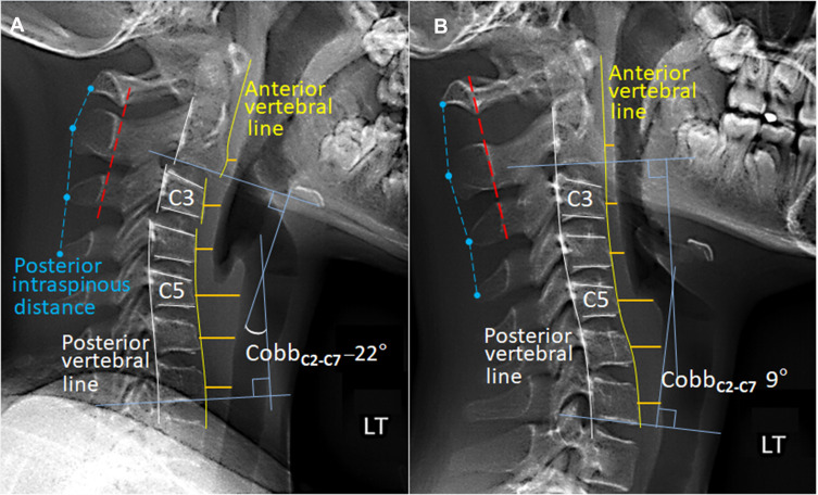
Comparison of cervical alignment on sagittal radiographs in neutral position. (A) At the initial assessment, several of the vertebrae were wedge-shaped, allowing for slippage of the C2 on C3 and C3 on C4.The global curvature of the C2-C7 spine using Cobb angle (four blue lines) was –22°, a reversed cervical lordosis. Intraspinous distance (dashed blue lines) was within 1.5 times the distance of the level either above or below and the spinous processes of C1 to C3 (dashed red line) was aligned within 1 mm of each other, suggestive an integrity of the interspinous ligaments. (B) 12-months later, the vertebral configuration became roughly rectangular. Restored cervical alignment and cervical lordosis yielded simultaneous remission of neurological symptoms. With reference to the initial image, there was 31° (–22° vs 9°) correction of the cervical lordosis. Reduced width of the prevertebral soft-tissue thickening (Orange lines) could signify a relief of previously swollen soft-tissue.