PMC full text:
Published online 2022 Sep 19.
Figure 3.
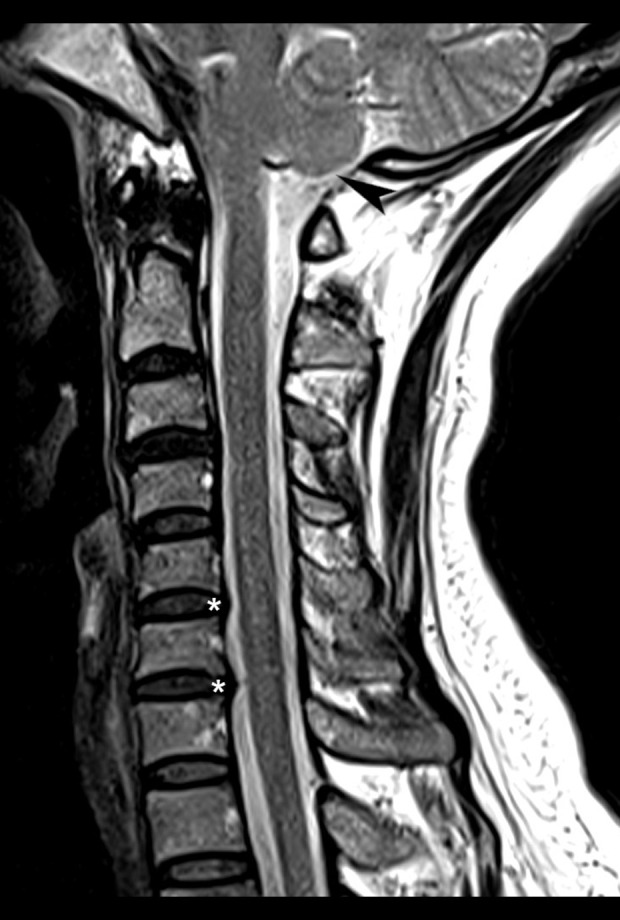
Cervical magnetic resonance imaging, T2-weighted sagittal view. There is evidence of spondylosis, including disc bulges (*), most evident in this view at C5–6 and C6–7. These were small, centrally located, and not contacting the spinal cord. There is no apparent central canal or neuroforaminal stenosis. Low-lying cerebellar tonsils are evident, measuring 0.39 cm inferior to the level of the foramen magnum (arrowhead).