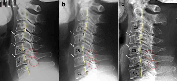
Figure 1

Radiography of a 44-year-old woman comparing the cervical alignment on the lateral view. (a) Lateral radiograph of the cervical spine at initial presentation shows grade 1 retrolisthesis with less than 25% posterior translation of C3 on C4 and C4 on C5 vertebrae (yellow lines highlight the posterior vertebral body margins). The inferior articular processes of C5 and C6 are lying on top of the superior articular processes of the below vertebrae (known as perched facets, highlighted by red circles). In addition, there is upper endplate sclerosis over the C4, C5, and C6 vertebrae (white arrows). (b) The radiograph obtained at the 4-year follow-up in 2013 demonstrates reduction of cervical retrolisthesis at all levels. (c) On the cervical radiograph taken in 2022, restoration of cervical alignment is observed after 13 years of manual maintenance care.