DOCUMENTATION OF COMPLIANCE MEASUREMENT
USED IN THE FORCE RECORDING AND ANALYSIS SYSTEM
J. M. Evans, C. L. Evans
Sense Technology, Inc.
BACKGROUND
Shortly after the introduction of the Model SHLCP-5
Precision Adjustor, reports were received from clinicians that the sound of the
impulse changed during use. Some clinicians began using the change in sound as
an indicator that the adjustment phase should be terminated.4 Another group of
clinicians5 began to use the instrument as a percussor to help locate points on
the spine as candidates for adjustment.
The use of percussive techniques
as an aid to diagnosis is well known in the medical arts. The image of the
physician tapping our body with a small hammer is a vivid childhood memory for
most.
As commonly practiced, this technique is subject to wide
variability in both the application of the percussive force (was the last
impulse the same as the current impulse?) and the interpretation of the results
(you can hear the difference, can't you?). This variability and the total
subjectivity of the interpretation of the results require a certain minimum
training and individual aptitude to make the technique useful and the results
transferable across patients and examiners.
The development of the Force
Recording and Analysis System presents an opportunity to improve the percussive
technique since:
- Each impulse on a given force setting is delivered with essentially the
same energy content.
- The peak force of the impulse is objectively measured and highly
repeatable when the impulse is delivered to a given substrate.
- Changes in the peak force of the impulse due to differences in the
substrate to which the impulse is applied are easily measured and objective.
With these potential benefits in mind, it was decided to
investigate the potential for improving the use of the Model SHLCP-5 Precision
Adjustor by incorporating an objective method of measuring substrate compliance
in the Force Recording and Analysis System Model 01.
PURPOSE
Development of a reliable objective method of assessing
differences in the response of the body to the application of a light impulse
loading.
THEORETICAL ANALYSIS
Impulse techniques are
commonly used in engineering analysis to examine the response characteristics of
structures or electrical circuits.6 7 These techniques employ the use of
instrumented impulse hammers to excite a structure and appropriate sensors to
measure the response of the structure. Of particular interest is the single
point vibration analysis in which the excitation and response of the structure
occur at the same point.
The adjusting head of the Force Recording and
Analysis System may be used in a manner consistent with single point
impulse-response testing. In this case the excitation of the "structure" is
achieved by the armature within the solenoid body being brought into contact
with the anvil of the adjusting head. To achieve this excitation, the adjusting
head must first be pressed against the patient until a pre-defined preload is
achieved. This assures that the initial conditions for the impulse are the same
each time.
At a given force level the energy imparted to the solenoid
armature is the same each time the head is activated. Since the mass of the
armature is also constant, the velocity of impact of the armature with the anvil
is essentially constant. The energy of excitation imparted to the anvil by the
armature will thus be constant each time the head is activated at a given force
level.
After the anvil has been excited by the armature, it is free to
move with respect to the adjusting head. The movement of the anvil against the
resistance of the substrate (patient simulator or patient body) determines the
force between the anvil and the patient's body as measured by the force
transducer attached to the front of the anvil. Under these conditions, "the
stiffness of the contacting surfaces affects the shape of the force
pulse..."8

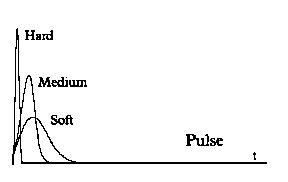
Illustration of Pulse Shapes Obtained From Different Substrates

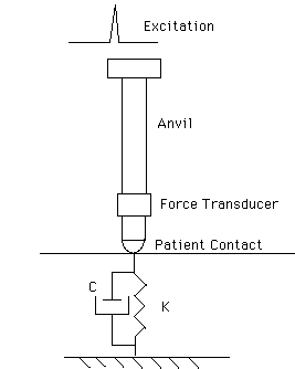
Illustration Using a Simple Discrete Parameter Model
The patient's body is represented by the spring with spring constant K and
the damper C. In the equation of motion for this system, the mass used would
include the mass of the anvil as well as a virtual mass of tissue. Assuming that
the damping and virtual mass are essentially constant, the frequency response of
the system and, therefore, the shape of the impulse would be determined
primarily by the spring constant or stiffness of the substrate.
If this
simple analysis is fundamentally sound, the peak force of each impulse would be
expected to vary with the stiffness (or inversely with the compliance) of the
substrate and would be expected to be constant at a point on a
substrate.
OBJECTIVE
Document rationale and performance of assessment
techniques.
MATERIALS
Sense Technology Force Recording and
Analysis System Model 01.
Patient simulators of various density and
elasticity.
Patient volunteer.
METHOD
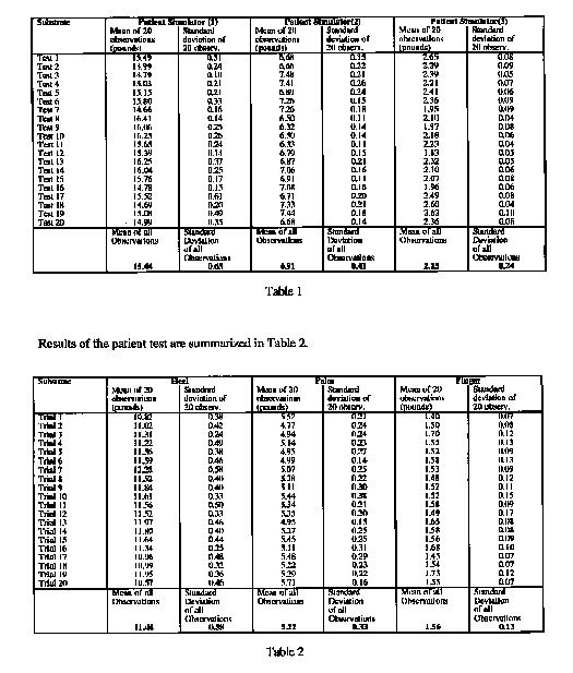
The adjusting
head of the Force Recording and Analysis System was placed on a substrate
simulating a point on the body of a patient and activated twenty times. The peak
force for each impulse was recorded. The mean peak force and standard deviation
of the observation was calculated. This test was repeated twenty times and the
standard deviation of the total data set was calculated. This is a measure of
the variability in the peak force measurements. Two peak force measurements many
standard deviations apart indicate a change in the physical system.
This
test was repeated on simulator materials of different compliance.
After
the tests were concluded on substrates of constant but different compliance, the
test was repeated on a volunteer. The first test point was chosen to be the heel
of the subject's hand, the second test point was the palm of the subject's hand
and the third was the tip of the index finger.
All tests were run at a
force setting of fifteen pounds and the peak force was recorded with the Force
Recording and Analysis System.
Results of the patient simulator tests are summarized in Table
1.
ANALYSIS
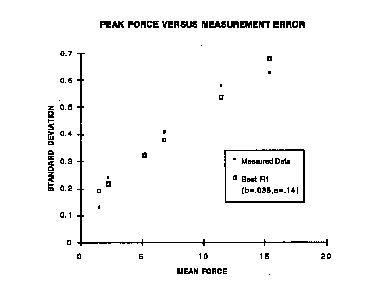
The results of the tests indicate that the peak force of
the impulse produced by the adjusting head of the Force Recording and Analysis
System can be reliably reproduced when the impulse is applied to a single point
on a unchanging substrate such as a patient simulator. The results also indicate
that the peak force of the impulse is repeatable when applied on one point of
the body. An estimation of the measurement error at each force level is shown in
the graph.
In addition, the mean force varies in a predictable and expected manner when
the impulse is applied to surfaces of differing compliance; that is, low
compliance substrates (high resistance) result in a higher peak impulse with
higher compliance substrates (lower resistance) substrates resulting in lower
peak forces. Furthermore, the variability of the data indicate that differences
in peak force of greater than ten percent (fifteen pounds at full scale) have a
ninety-five percent chance of representing valid differences in the underlying
substrate.
CONCLUSIONS
The results of the testing and
analysis indicate that fundamental engineering analysis methodologies may be
productively applied to the problem of quantifying the analysis of the
compliance of the human body. In particular, single point vibration analysis
techniques utilizing impulse loading are useful in explaining and analyzing the
force output of the adjusting head of the Sense Technology Force Recording and
Analysis System. The peak force has been shown to be related to the compliance
of the substrate to which the adjusting head is applied. The peak force has been
shown to be essentially constant for a single substrate. Variations of greater
than ten percent in the peak force indicate a high probability that the output
was derived from two different substrates.
APPENDIX II Intra- and
Inter-Examiner Reliability Studies
DOCUMENTATION OF INTRA- AND INTER-EXAMINER
RELIABILITY OF
DIFFERENTIAL COMPLIANCE METHODOLOGY
(a pilot
study)
K. Allen D.C., R. Crisman, D.C., R. Keeler, D.C.,
J. Pesce, D.C. S.
Saleeby, D.C., J. M. Evans, Ph.D.
The compliance of the human spine may be thought of as the ease of movement
of each individual vertebra. For the purposes of this paper, compliance is
defined as the displacement response of a structure when subjected to a unit
force. It is the inverse of stiffness and intuitively can be thought of as the
flexibility of a structure. This paper describes reliability studies on an
instrument which measures the compliance of the human spine, before, during and
after adjustment.
Sense Technology, Inc. has developed a unique
chiropractic adjusting system which incorporates a percussive adjusting head.
This percussor is instrumented with a force transducer that supplies data to a
computer system. The computer stores and displays the force data for clinical
evaluation. This system, referred to as the Force Recording and Analysis System
(FRAS), is an extension of previous adjustors which are marketed without the
force instrumentation.9 The clinician uses the Force Recording and Analysis
System to challenge each vertebra with a low energy impulse. The system records
and displays the peak force measured at each vertebra. The compliance of the
vertebra is inversely proportional to the peak force. The results of the
compliance assessment are used by the clinician, in conjunction with other
diagnostic techniques (such as palpation, X-ray examination, thermal analysis,
and analysis of patient complaint and history) to determine appropriate
adjustment locations.
BACKGROUND
Sigler and Howe [20] and
Jackson et al. [10] have pointed out that when measurement systems are used as
the basis for diagnostic techniques, the reliability of the measurement system
directly influences the reliability of the diagnostic system. Sigler and Howe
found that the errors involved in measuring very small changes in atlas position
on X-ray films were of such magnitude that the validity of the entire diagnostic
and therapeutic regime was open to question. Jackson et. al. found using a
somewhat different measurement system that changes in angles between vertebrae
could be reliably obtained from X-ray films. A series of studies has been
conducted examining the reliability of palpation and motion palpation for the
detection of "somatic dysfunction" with mixed results [4,14,15,17,25,27]. Where
statistical significance has been achieved, the level or strength of the
findings has been relatively low. DeBoer et. al. [4] points out that such
findings are not surprising since even well-established procedures such as
reading X-rays, E K G's or blood pressure give maximum intra-examiner
correlation in the range of .40 to .60. Others [19,21-24] have attempted to
assess the reliability of simple instrumentation (inclinometers, temperature
measurement instruments and penetration devices) as aids to diagnosis, again
with mixed results. Several authors [7,8,16] have critiqued the methodology used
in intra- and inter- examiner reliability studies. Their critiques may be useful
as general guides and to raise questions regarding statistical
methodology.
In previous work[6] we have documented the reliability and
applicability of force measurement techniques for measuring differences in
compliance of various substrates including the human body. That study concluded
that the peak force of the impulse of the adjusting head varies directly with
the stiffness of the substrate ( inversely with the compliance). The measures
obtained showed that the system produced a repeatable result and that
differences of greater than ten percent in peak force were
significant.
Clinicians are currently using the Model SHLCP-5 Precision
Adjustor to percussively test the spine both before and after adjustment [3].
The force levels used are generally higher (twenty-five pounds) than used for
the initial assessment of the Force Recording and Analysis System (fifteen
pounds). The proposal to apply these techniques to the analysis of compliance
along the human spine raises some fundamental questions. This study examined the
following:
- Will the measured peak forces resulting from application of very light
impulse loads differ significantly vertebra to vertebra?
- How will differences in patient physique (including body fat content,
muscle size, muscle tone, etc.) affect the results?
- Will the pattern of force readings be reproducible on a patient?
- Will the pattern of force readings be reproducible by more than one
examiner?
In order to provide preliminary answers to these
questions, the following trials were conducted:
Trial One- Examine the
variability of compliance measurements in the cervical spine
Trial Two-
Determine the reproducibility of repeated compliance readings taken by the same
examiner.
Trial Three- Determine the reproducibility of repeated
compliance measures taken by two different examiners.
TRIAL ONE
Determine the variability of compliance measurements
of the human
spine using the Force Recording and Analysis System.
Method: A 30mm dual
prong attachment was used to straddle the
spinous of the patient's vertebrae.
The examining chiropractor
stabilized the head of the seated patient in
flexion, chose the line
of drive and positioned the tips of the attachment on
the
appropriate vertebra. Measurements were taken in the cervical
area of
the spine of ten patients.
Results: Variations of peak force of up to 55%
were found along the cervical
spine of individual patients.
Analysis:
Some variation in measured peak force along the spine would be
expected from
measurement variability alone. The commonly
observed differences in peak
force of over ten percent between
adjacent vertebrae suggest that there is
information in the
measurements that cannot be explained by measurement
variability [6]. This is suggestive of clinical
significance.
Occasional differences in the mean of the cervical
measurements
between patients were observed during the initial testing
phase.
These differences were eliminated by normalizing each set of
measurements against the highest reading before display. The
normalized
display showed the peak force readings as a percent of
the highest peak
force reading for each patient. These normalized
displays allow the
clinician to focus on the difference between
peak force readings rather than
their absolute value.

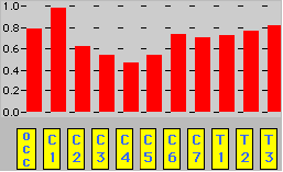
Illustration of Normalized Display
TRIAL TWO
Determine the reproducibility of repeated compliance
readings
taken by the same examiner.
Method: Using the 30mm dual prong
attachment to straddle the
spinous of the patient's cervical vertebrae, the
examining
chiropractor stabilized the head and neck of the seated
patient
in flexion. The examining chiropractor chose the line of drive
and
positioned the tips of the instrument on the occiput, and
vertebrae C1-7 and
T1-3. Twenty patients participated in the study.
The examiner obtained a
second set of readings on each patient
immediately after the first set. There
were no markings on the
patient to serve as position references.
Research
Hypothesis: The patterns of compliance measurements
obtained using the
Force Recording and Analysis System in consecutive
measure-
ments of the human cervical spine by the same examiner show
no
significant differences from one trial to the
next.
Statistical
Analysis: Each set of consecutive readings was
examined for similarity
using the following statistical techniques. First,
the probability that
the two sets of data were different was computed using
a c2
statistic. This method was chosen since an estimate of the
measurement error at each force level was available.[6] The c2
statistic
was computed using the formula:
where: xi = the first set of data
collected from the patient
yi = the second set of data
= the standard
deviation at the force level
= the standard deviation at the force level
and are calculated using the empirical relationship
developed in
[6]:
where: = .14
= .035
The probability of obtaining a c2
value at least as large as that
observed, assuming the measurements were
drawn from the same
distribution, was computed from an incomplete gamma
function.10
If this probability is smaller than .05, then the hypothesis
that the
data sets are the same may be rejected with a confidence of
95%.
In addition, the Pearson product moment correlation (r ) was
computed on the data sets using the formula:
where: xi = the
first set of data collected from the patient
yi = the second set of
data
Finally, the intra-class correlation coefficient (ICC) was computed
by performing a one way analysis of variance and formulating the
ICC
according to:
where = the variance within the data
= the
variance between the data sets
m = the number of levels of the
analysis
The chi square metric computes the square of the difference
between each paired observation (the value obtained at level C1 on the first
observation is subtracted from the value obtained at C1 on the second
observation and the difference squared) and then compares that value to an
estimate of the measurement, determined through empirical repetitive testing on
known substrates of constant compliance[6].
If the measurements are
exactly the same, the chi square returns a value of zero and the probability of
obtaining a poorer (larger) result is necessarily equal to one. The larger the
chi square the less likely that the two sets of measurements are "the same" or
that the measurement is repeatable within an acceptable degree of
error.
The chi square is a good measure of the significance of the
difference between two sets of measures. Because its numeric values are not
subject to direct interpretation, it does not provide a measure of the strength
of the association. Some form of correlation coefficient is generally used as a
measure of the strength of a significant association. The most widely used is
the linear correlation (also referred to as the product-moment correlation or
Pearson's r). The use of this statistic has been criticized [7,8,16], primarily
because of artifacts such as obtaining a high correlation even though the two
sets of measures differed by some constant value (i.e., a correlation equal to
one would be obtained from two data sets even though each value in the second
data set were twice the value of its mate in the first data set). In our case,
where the pattern of data within the set may be more important than the exact
values, this limitation may not be important.
The intra-class
correlation coefficient (ICC) has been proposed, [7,16] as a more reliable
indicator of the strength of a significant association for reliability studies
involving continuous measurements. This coefficient can be constructed from the
results of a one way analysis of variance. The formulation
is:
where = the variance within the data
= the variance
between the data sets
m = the number of levels of the analysis
If the
data sets are exactly the same then MSB equals zero and the ICC equals one. If
the variance between the data sets is greater than the variance within the data
set then the ICC will be negative (no agreement between levels). If the variance
between the data sets is less than the variance within the data then the ICC
will be positive and there is said to be some agreement between levels. The
results are summarized in TABLE 1.
Examiner Patient p r
1 1 10.52 .484
.98
1 2 41.23 <.0000 .86
1 3 12.01 .363 .85
1 4 11.94 .368 .80
1
5 12.62 .319 .85
1 6 9.47 .579 .80
1 7 10.89 .452 .82
1 8 12.11 .355
.74
1 9 3.73 .977 .97
1 10 15.56 .158 .82
1 11 14.82 .191 .80
1 12
11.5 .402 .84
1 13 20.61 .038 .91
1 14 15.68 .153 .82
1 15 15.35 .167
.86
Linear Correlation Across Patients for Examiner One =.88
ICC
Across Patients for Examiner One =.90
2 1 18.58 .069 .89
2 2 9.21 .602
.95
2 3 14.43 .210 .91
2 4 49.91 <0000 .72
2 5 19.94 .046
.86
Linear Correlation Across Patients for Examiner Two =.85
ICC
Across Patients for Examiner Two =.93
Linear Correlation Across Patients
for Examiner One and Two =.96
ICC Across Patients for Examiner One and Two
=.96
TABLE 1- INTRA-EXAMINER RELIABILITY

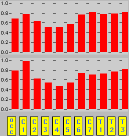
Example of results judged to be "different" according to the c2
criterion.

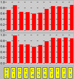
Example of results judged to be "the same" according to the c2
criterion.
Analysis: The two consecutive readings taken in the cervical area
were
highly correlated for each chiropractor. In addition,
the c2 probability
indicates that the null hypothesis (the
measurements are the same) could be
rejected in only three
cases. It appears that the intra-examiner
reliability
of the tests is high, by any of the metrics. The chi-squared and
ICC metrics sometimes disagree since chi-squared normalizes the
observed
variability against an empirical estimate of the
measurement error, and the
ICC normalizes against the variability
in the patient's measurements. If the
ICC is calculated for all of the
data, a very high value is returned. This is
done in the
overall ICC measurements reported in TABLE 1
above.
Sources of Variability:
There are two obvious sources of
variability which may influence
the outcome of this intra-examiner
reliability study. The first is
the error introduced due to imperfect
placement. By placement,
we mean the position of the dual prong tips used for
contacting
the patient during the examination as well as the line of drive
chosen by the examiner. Even with markings on the cervical area
(which
were not used in this study), the angle of the instrument
and the positioning
of the tips would be impossible to duplicate
exactly from the first
examination to the second.
Another source of error is a result of the
measurement itself.
In our case, it is not difficult to understand that the
second
set of measurements may differ from the first due to the act of
measuring because the energy used in the measurement may
well be
sufficient to cause changes in the underlying structure
of the spine. Such
changes would be expected to result in
differences in response to the energy
of the test impulse. That
these changes are relatively small is attested to
by the excellent
agreement found. This agreement may well be improved
by
training and/or lowering the energy of the impulse.
Conclusions:
The intra-examiner reliability of compliance measurements
obtained in the
cervical spine with the Force Recording and
Analysis System is consistently
high.
TRIAL THREE
Determine the reproducibility of repeated
compliance readings
taken by two different examiners.
Method: Using
the 30mm dual prong attachment to straddle the spinous
of the patient's
cervical vertebrae, an examining chiropractor
stabilized the head and neck of
the seated patient in flexion.
The first examining chiropractor chose the
line of drive and
positioned the tips of the instrument on the occiput
and
vertebrae C1-7 and T1-3. Three patients participated in the study.
A
second examiner obtained a second set of readings on each patient
immediately
after the first set. There were no markings on the
patient to serve as
position references.
Research
Hypothesis: The patterns of compliance
measurements obtained using
the Force Recording and Analysis System in
consecutive
measurements of the human cervical spine by two
different
examiners show no significant differences from one trial to
the
next.
Statistical
Analysis: This analysis was conducted in the same
manner as the
previous analysis for the intra-examiner reliability.
Examiner Patient p r
1-3 21 10.95 .447 .80
1-3 22 8.45 .673
.87
1-3 23 11.4 .432 .75
Linear Correlation Across Patients for
Examiner One and Three =.89
ICC Across Patients for Examiner One and Three
=.65
Analysis: The two consecutive readings taken in the cervical
area
were highly correlated for each chiropractor. In addition,
the c2
probability indicates that the null hypothesis (the
measurements are the
same) could not be rejected in any
of the cases. It appears that the
intra-examiner repeatability
of the tests is high.
Discussion: The
purpose of this trial is to determine whether
or not the compliance
measurements obtained by two different
clinicians on a single patient are
the same. The statistics suggest
that the measurements are quite
reproducible across clinicians.
CONCLUSION
This study
suggests that the measurements of the FRAS system reflect the actual compliance
of the patient's spine. Further, the intra- and inter- examiner reproducibility
of these measurements is good. A subsequent study will quantify our observations
that these compliance measurements change after chiropractic adjustment with the
instrument. Clinicians are just beginning to develop diagnostic and treatment
rules to use the information provided by this new instrument.
Sense
Technology Inc.
12/15/94
NOTES ON THE VALIDITY OF COMPLIANCE
MEASUREMENTS OBTAINED WITH THE FORCE RECORDING AND ANALYSIS SYSTEM
Rob Crisman DC
ABSTRACT
OBJECTIVE
To verify that striated muscle contraction
reduces the response of the skeletal structure to a low energy impulse when
compared to the relaxed state. In addition, to verify that joints of different
mobility will have predictable outcomes, i.e., when tested with a low energy
impulse, skeletal joints with low mobility will elicit a higher response than
skeletal joints with higher mobility.
DESIGN
Four normal volunteers
were recruited to investigate the effects of muscle spasm on the readings
obtained with the Force Recording and Analysis System (FRAS). Additionally a
series of joints with known and different gross compliance were tested.
SETTING
Private clinical practice.
INTERVENTIONS
None.
MAIN OUTCOME MEASURES
Differential Compliance readings obtained with
the Force Recording and Analysis System.
RESULTS
The results
verified that the measurements taken with the FRAS on human subjects confirmed
the hypotheses postulated by the investigator that muscle spasm may produce
lower readings and that measurements taken on joints of low compliance versus
joints of high compliance in human volunteers varied in the manner expected.
CONCLUSION
Both hypotheses were confirmed by the tests. It appears
that the compliance measurements obtained using the FRAS vary predictably in the
expected manner and may be useful in the clinical setting.
KEY INDEXING
TERMS
CFI, Computer, Fixation, Medical Imaging, Compliance.
INTRODUCTION
The Force Recording and Analysis System (FRAS)
manufactured by Sense Technology Inc. is used to perform a Computerized Fixation
Imaging Scan (CFI Scan) of a patient's skeletal structure. The Differential
Compliance Methodology implemented with the FRAS enables the examiner to
objectively determine the relative compliance of two or more vertebral segments
or to evaluate the mobility of a single spinal segment in any of its degrees of
freedom.
The first step in obtaining a CFI Scan is to obtain a measure
of the compliance of each vertebra or segment of the spine under evaluation.
This measure is obtained by applying a low energy mechanical impulse to each
segment of the spine sequentially and recording the response of the segment.
After all segments of interest have been challenged in this way, the responses
are displayed visually for the examiner as a bar graph. Low bar graph readings
indicate a segment with high compliance (low resistance) and high bar graph
readings indicate a segment of low compliance (high resistance). The clinical
interpretation of these results is that a joint segment with a much lower
compliance than its neighbors may be fixated and a candidate for adjustment.
Previous work1 has demonstrated that the FRAS instrument gives reliable
and predictable results when applied to surfaces of known and constant
compliance and to parts of the body with different compliance. Clinical use of
the instrument, while supporting the general conclusions drawn from these tests,
raises questions regarding the role of the musculature surrounding and attached
to skeletal segments. In particular, striated muscle contraction surrounding a
joint segment commonly found in soft tissue injury2.may influence the results of
the scan. If the muscle contraction reduces the normal response of the segment,
a joint segment that is in fact fixated may be judged normal or of high
compliance.
The purpose of this study is to examine the hypothesis that
muscle contraction reduces the response of a skeletal segment to the mechanical
impulse used to challenge the segment. In addition, a series of joints of
different compliance were tested to examine the hypothesis that joints of low
compliance produce higher readings when compared to joints of high compliance.
METHODS
SUBJECTS
Four normal, pain free volunteers (two
females: the first, 26 years of age, 170 cm in height, 66 kg in weight; the
second, 33 years of age, 165 cm in height, 65.8 kg in weight; and two males: the
first, 27 years of age, 180 cm in height; 86 kg in weight; the second, 32 years
of age, 183 cm in height, 88.4 kg in weight) were recruited for the study. Each
subject was required to read an information sheet describing the study and give
written informed consent before being included in the study.
PROCEDURE
EFFECT OF MUSCLE SPASMS
In order to test the effect
of muscle spasm or tetanus on the compliance readings obtained with the Force
Recording and Analysis System, one site on each subject was tested. The site
chosen was the anterior midpoint of the diaphysis of the right humerus. The
midpoint was visually determined after palpation of both ends of the bone. This
point, coincident with the belly of the bicep muscle, is the thickest part of
the muscle when in full contraction. Tests were made with the elbow bent at
approximately 35 degrees, 90 degrees and 150 degrees with the bicep muscle in
full isotonic contraction. The same point was tested three times at 35 degrees,
three times at 90 degrees, twice at 150 degrees and three times with no
contraction.
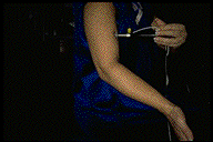
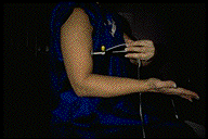

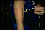
For convenience, the tests were conducted using an
eleven point data collection and analysis algorithm. In general, the first eight
readings were obtained with the muscle under examination in a contracted state.
Then, without leaving the data collection routine, the last three readings were
obtained with the muscle in a relaxed normal state of tetany. After all eleven
data points were collected, the algorithm normalized the results and displayed
all the data points as a series of eleven bars in graph format.
COMPARISON OF JOINTS OF DIFFERENT COMPLIANCE
In order to compare
compliance measurements obtained on joints of known and different compliance, a
series of tests were made on sutura, condyloid and ginglymus joints. These tests
were conducted on the male subjects The tests were conducted in the presumed
order of lowest compliance to the highest compliance, i.e. the sutura was tested
first followed by the condyloid and ginglymus joints. The specific joints chosen
were:
- Sutura: left temporal/parietal joint
- Condyloid: right trapezoid bone
- Ginglymus: second digit- middle phalangeal
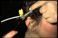
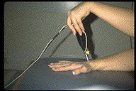
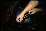
Again, for convenience, an eleven point algorithm
was used for data collection and analysis. Each joint with the exception of the
last was challenged four times with the low force impulse and the response
recorded. The last joint was challenged three times.
DATA ANALYSIS
Each skeletal segment of interest is challenged by a low energy impulse.
This impulse contains a wide band of frequencies (from zero to approximately
20,000 Hertz). The segment resists the initial impulse and the amount of
resistance is recorded with a force transducer. The peak force recorded is a
simple measure of the resistance of the segment to the initial low energy
impulse. More complex analysis of the response waveform would reveal the major
frequency of the response of the segment. For our purposes, the maximum response
was sufficient to characterize the compliance of each of the skeletal segments
of interest.
After the segment has been challenged and the maximum
response recorded, the analysis routine asks for the next segment. The
investigator repeats the test on all segments of interest in turn. In this case
the analysis routine was composed of eleven segments. After all eleven segments
have been tested, the analysis routine selects the segment with the maximum
response and sets its value to one. The values of the remaining segments are
transformed to a value proportional to the maximum value by the following
relationship:
fn = fi /fmax
where fn = the normalized maximum value for segment i
fi = the maximum value for segment i
fmax = the maximum value recorded
for all segments
This yields a set of values for the segments tested that are normalized
relative to the maximum value recorded. These values are then displayed for the
investigator as a bar graph with eleven elements.
RESULTS
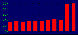
Typical results
obtained from the humerus with the elbow bent at 35 degrees, 90 degrees, and 150
degrees and relaxed. First nine measurements with muscle in full isotonic
contraction with the elbow at the three respective angles; first three
measurements at 35 degrees, second three at 90 degrees, next three at 150
degrees, remaining measurements with muscle relaxed.
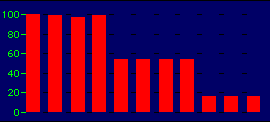
Typical results obtained from sequential testing of sutura joint, condyloid
joint, and ginglymus joint. First four measurements obtained from the sutura
joint; second four measurements from the condyloid; last three measurements from
the ginglymus.
DISCUSSION
The results of the tests clearly indicate that
contracted versus relaxed striated muscle gives lower readings when the muscle
is contracted. The reason for this result appears to be the greater absorption
of the energy of the test impulse when the test is conducted on contracted
striated muscle. The greater absorption may be due to the actin and myosin
filament crossing over each other in the sacromere, causing the muscle to be
thicker at the belly of the muscle.3 Also, the tension of the muscle creates a
spring board effect which, when combined with the damping properties of tissue,
acts to shield the underlying structure much like a spring and shock absorber in
an automobile. Conversely, when the muscle was relaxed, the impulse penetrates
directly to the underlying bone which produces a higher response to the impulse.
When a low reading is obtained clinically with the FRAS, the clinician
must determine whether the reading represents a highly mobile (hypermobile?)
segment or whether the reading is due to muscular involvement. This
determination is relatively straightforward and is made primarily through
palpation of the segment in question to determine evidence of tenderness or
muscle tightness.
CONCLUSION
Two hypothesis were postulated by the examiner. The
first was that a joint surrounded by muscle in contraction would produce a low
response to a low energy mechanical impulse when compared to the response to the
same impulse when the muscle is not in contraction. The hypothesis was tested by
challenging the skeletal body at one point where the contraction of the
overlying muscle was easily controlled. The point was the belly of the bicep
muscle over the midpoint of the humerus. The intensity of the contraction was
controlled by varying the angle between the humerus and the forearm. Observation
of the test results showed that the more intense the contraction at either site
correlated with lower readings (higher apparent compliance). These results
supported the hypothesis and confirmed instructions in the manufacturer's
manuals as well as the clinical protocol co-authored by the author.
The
second hypothesis was that joints with different compliance would produce
different results when tested and that the results would vary predictably with
known joint compliance when compared on the same scale. This hypothesis was also
confirmed. The response obtained from the sutura was 40 percent higher than the
responses obtained from the condyloid and approximately 500 percent greater than
that obtained from the ginglymus.
Both hypotheses were confirmed by the
tests. It appears that the compliance measurements obtained using the FRAS vary
predictably in the expected manner and may be useful in the clinical setting.
REFERENCES
1. Evans, J M, Evans, C L. Documentation of Compliance
Measurement Used in the Force Recording and Analysis System, Sense Technology
Inc., 1994
2 Gordon, Huxley and Julian, "The Length-tension Diagram of
Single Vertebrate Striate Muscle Fibers, "Journal of Physiology 171:28P,
1964.
3 Lan, N.C. et al., "Mechanisms of Glucocorticoid Hormone
Action,"Journal of Steroid Biochemistry, 20:77, 1984.
Take Me
Home


![]()


![]()

![]()


