A Patient's Guide to Cervical Radiculopathy
Introduction
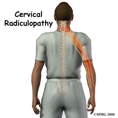
Neck pain has many causes. Mechanical neck pain comes
from injury or inflammation in the soft tissues of the neck.
This is much different and less concerning than symptoms
that come from pressure on the nerve roots as they exit the
spinal column. People sometimes refer to this problem as a
pinched nerve. Health care providers call it
cervical radiculopathy.
This guide will help you understand
- how the problem develops
- how doctors diagnose the condition
- what treatment options are available
Anatomy
What part of the neck is involved?
The spine is made of a column of bones. Each bone, or
vertebra, is formed by a round block of bone, called
a vertebral body. A bony ring attaches to the back of
the vertebral body. When the vertebra bones are stacked on
top of each other, the bony rings forms a long bony tube
that surrounds and protects the spinal cord as it passes
through the spine.
Travelling from the brain down through the spinal column,
the spinal cord sends out nerve branches through openings on
both sides of each vertebra. These openings are called the
neural foramina. (The term used to describe a single
opening is foramen.)
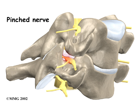
The intervertebral disc sits directly in front of
the opening. A bulged or herniated disc can narrow the opening and put pressure
on the nerve. A facet joint sits in back of the
foramen. Bone spurs that form on the facet joint can project
into the tunnel, narrowing the hole and pinching the
nerve.
An intervertebral disc fits between the vertebral bodies
and provides a space between the spine bones. The disc
normally works like a shock absorber. An intervertebral disc
is made of two parts. The center, called the nucleus,
is spongy. It provides most of the shock absorption. The
nucleus is held in place by the annulus, a series of
strong ligament rings surrounding it. Ligaments are
strong connective tissues that attach bones to other
bones.
Related Document: A
Patient's Guide to Cervical Spine Anatomy
Causes
Why do I have this problem?
Cervical radiculopathy is caused by any condition that
puts pressure on the nerves where they leave the spinal
column. This is much different than mechanical neck pain.
Mechanical neck pain is caused by injury or inflammation in
the soft tissues of the neck, such as the discs, facet
joints, ligaments, or muscles.
The main causes of cervical radiculopathy include
degeneration, disc herniation, and spinal instability.
Degeneration
As the spine ages, several changes occur in the bones and soft tissues. The disc
loses its water content and begins to collapse, causing the
space between the vertebrae to narrow. The added pressure
may irritate and inflame the facet joints, causing them to
become enlarged. When this happens, the enlarged joints can
press against the nerves going to the arm as they try to
squeeze through the neural foramina. Degeneration can also
cause bone spurs to develop. Bone spurs may put pressure on
nerves and produce symptoms of cervical radiculopathy.
Herniated Disc
Heavy, repetitive bending, twisting, and lifting can
place extra pressure on the shock-absorbing nucleus of the
disc. A blow to the head and neck can also cause extra
pressure on the nucleus. If great enough, this increased
pressure can injure the annulus (the tough, outer ring of
the disc). If the annulus ruptures, or tears, the material
in the nucleus can squeeze out of the disc. This is called a
herniation.
Although daily activities may cause the nucleus to press
against the annulus, the body is normally able to withstand
these pressures. However, as the annulus ages, it tends to
crack and tear. It is repaired with scar tissue. Over time,
the annulus becomes weakened, and the disc can more easily
herniate through the damaged annulus. If the herniated disc
material presses against a nerve root it can cause pain,
numbness, and weakness in the area the nerve supplies.
Spinal Instability
Spinal instability means there is extra movement among
the bones of the spine. Instability in the cervical
spine (the neck) can develop if the supporting ligaments
have been stretched or torn from a severe injury to the head
or neck. People with diseases that loosen their connective
tissue may also have spinal instability. Spinal instability
also includes conditions in which a vertebral body slips
over the one just below it. When the vertebral body slips
too far forward, the condition is called
spondylolisthesis. Whatever the cause, extra movement
in the bones of the spine can irritate or put pressure on
the nerves of the neck, causing symptoms of cervical
radiculopathy.
Symptoms
What does the condition feel like?
The symptoms from cervical radiculopathy are from
pressure on an irritated nerve. These symptoms are not the
same as those that come from mechanical neck pain.
Mechanical neck pain usually starts in the neck and may
spread to include the upper back or shoulder. It rarely
extends below the shoulder. Headaches are also a common
complaint of both radiculopathy and mechanical neck
pain.
The pain from cervical radiculopathy usually spreads
further down the arm than mechanical neck pain. And unlike
mechanical pain, radiculopathy also usually involves other
changes in how the nerves work such as numbness, tingling,
and weakness in the muscles of the shoulder, arm, or hand.
With cervical radiculopathy, the reflexes in the muscles of
the upper arm are usually affected. This is why doctors
check reflexes when people have symptoms of cervical
radiculopathy.
Diagnosis
How do doctors diagnose the problem?
Doctors gather the information about your symptoms as a
way to determine which nerve is having problems. Diagnosis
begins with a complete history of the problem. Your doctor
will ask questions about your symptoms and how your problem
is affecting your daily activities. Your answers can help
your doctor determine which nerve is causing problems.
Next, the doctor examines you to see which neck movements
cause pain or other symptoms. Your skin sensation, muscle
strength, and reflexes are tested in order to tell where the
nerve problem is coming from.
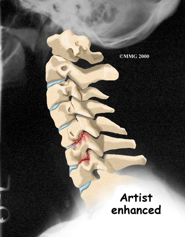
X-rays of the cervical spine can show the cause of
pressure on the nerve. The images show whether degeneration
has caused the space between the vertebrae to collapse. They
may also show if a bone spur is pressing against a
nerve.
If more information is needed, your doctor may order
magnetic resonance imaging (MRI). The MRI machine
uses magnetic waves rather than X-rays to show the soft
tissues of the body. This test gives a clear picture of the
discs, nerves, and other soft tissues in the neck. The
machine creates pictures that look like slices of the area
your doctor is interested in. The test does not require any
special dye or needles and is painless.
Sometimes it isn't clear where the nerve pressure is
coming from. Symptoms of numbness or weakness can also
happen when the nerve is being pinched or injured at other
points along its path. (An example of this is pressure on
the median nerve in the wrist, known as carpal tunnel
syndrome.) Electrical studies of the nerves going from
the neck to the arm may be requested by your doctor to see
whether the nerve problem is in the neck or further down the
arm. However, most doctors take X-rays and try other forms
of treatment before ordering electrical tests. These tests
are usually only needed when the diagnosis is not clear.
If your doctor orders electrical studies, several tests
are available to see how well the nerves are functioning,
including the electromyography (EMG) test. This test measures
how long it takes a muscle to work once a nerve signals it
to move. The time it takes will be slower if nerve pressure
from radiculopathy has affected the strength of the
muscle.
Another electrical test that may be used instead of EMG
is cervical root stimulation (CRS). This test
involves putting a small needle through the back of the neck
into the nerve where it leaves the spinal column. Readings
of muscle action are then taken of the muscles on the front
and back of the upper arm and along the inside of the lower
arm. Doctors use the readings to determine which nerve is
having problems.
Treatment
What treatment options are available?
Nonsurgical Treatment
Unless the nerve problem is getting worse rapidly, most
doctors will begin with nonsurgical treatments.
At first, your doctor may prescribe immobilization of the
neck. Keeping the neck still for a short time can calm
inflammation and pain. This might include one to two days of
bed rest and the use of a soft neck collar. This collar is a padded ring that
wraps around the neck and is held in place by a Velcro
strap. Normally, a patient need only wear a collar for one
to two weeks. Wearing it longer tends to weaken the neck
muscles.
Doctors prescribe certain types of medication for
patients with cervical radiculopathy. Severe symptoms may be
treated with narcotic drugs, such as codeine or morphine.
But these drugs should only be used for the first few days
or weeks after problems with radiculopathy start because
they are addictive when used too much or improperly. Muscle
relaxants may be prescribed to calm neck muscles that are in
spasm. You may be prescribed anti-inflammatory medications
such as aspirin or ibuprofen. Some doctors have their
patients work with a physical therapist. At first,
treatments are used to ease pain and inflammation.
Electrical stimulation treatments can help calm muscle spasm
and pain. Traction is a way to gently stretch the joints and
muscles of the neck. It can be done using a machine with a
special head halter, or the therapist can apply the
traction pull by hand.
Some patients are given an epidural steroid injection (ESI). The spinal
cord travels in a tube within the bones of the spinal canal.
The cord is covered by a material called dura. The
space between the dura and the spinal column is the
epidural space. It is thought that injecting steroid
medication into this space fights inflammation around the
nerves, the discs, and the facet joints. In some cases, the
steroid injection is given around one specific nerve. This
is called a selective nerve block. The response to
this treatment helps confirm which nerve root is causing the
symptoms.
Doctors usually have their patients try nonoperative
treatments for at least three months before considering
surgery. But when patients simply aren't getting better, or
if the problem is becoming more severe, surgery may be
suggested.
Surgery
Most people with cervical radiculopathy get better
without surgery. In rare cases, people don't get relief with
nonsurgical treatments. They may require surgery. There are
several types of surgery for cervical radiculopathy. These
include
- foraminotomy
- discectomy
- fusion
Foraminotomy
A foraminotomy is done to open the neural foramen
and relieve pressure on the spinal nerve root. A foraminotomy may be done because of bone spurs or
inflammation.
Related Document: A
Patient's Guide to Cervical Foraminotomy
Discectomy
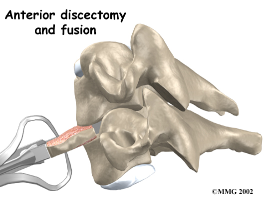
In a discectomy, the surgeon removes the disc
where it is pressing against a nerve. Surgeons usually
perform this surgery from the front (anterior)of the
neck. This procedure is called anterior cervical discectomy. In most patients,
discectomy is done together with a procedure called
cervical fusion, which is described next.
Related Document: A
Patient's Guide to Cervical Discectomy
Fusion
A fusion surgery joins two or more bones into one solid
bone. The purpose for treating cervical radiculopathy with
fusion is to increase the space between the vertebrae,
taking pressure off the nerve. The surgery is most often
done through the front of the neck. After taking out the
disc (discectomy), the disc space is filled in with a small
block of bone graft. The bone is allowed to heal, fusing the
two vertebrae into one solid bone. The space between the
vertebrae is propped and held open by the bone graft, which
enlarges the neural foramina, taking pressure off the nerve
roots.
Related Document: A
Patient's Guide to Anterior Cervical Discectomy and
Fusion
Rehabilitation
Nonsurgical Rehabilitation
What should I expect from treatment?
Even if you don't need surgery, your doctor may recommend
that you work with a physical therapist. Patients are
normally seen a few times each week for one to two months.
In severe cases, patients may need a few additional weeks of
care.
Your therapist creates a program to help you regain neck
and arm function. Treatments for cervical radiculopathy
often include neck traction, described earlier. Though neck
traction is often done in the clinic, your therapist may
give you a traction device to use at home.
It is very important to improve the strength and
coordination in the neck and shoulder blade muscles. Your
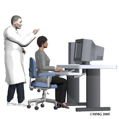
therapist can also evaluate your workstation or the way you
use your body when you do your activities and suggest
changes to avoid further problems.
After Surgery
Rehabilitation after surgery for cervical radiculopathy
can be a slow process. You will probably need to attend
physical therapy sessions for six to eight weeks, and you
should expect full recovery to take up to four months.
During physical therapy after surgery, your therapist may
use treatments such as heat or ice, electrical stimulation,
massage, and ultrasound to help calm pain and muscle spasm.
Then you begin learning how to move safely with the least
strain on the healing neck.
As the rehabilitation program evolves, you will do more
challenging exercises. The goal is to safely advance your
strength and function. As your therapy sessions come to an
end, your therapist will help you with decisions about
getting you back to work. Your therapist can do a work
assessment to make sure you'll be able to do your job
safely. Your therapist may suggest changes that could help
you work safely, with less chance of reinjuring your
neck.
When your treatment is well under way, regular visits to
the therapist's office will end. The therapist will continue
to be a resource for you. But you will be in charge of doing
your exercises as part of an ongoing home program. | 