PRACTICE ESSENTIALS
Patellofemoral Syndrome
Diagnostic Pointers and Individualized Treatment
Michele LaBotz, MD
Practice Essentials Series Editors:
Kimberly G. Harmon, MD;
Aaron Rubin, MD
THE PHYSICIAN AND SPORTSMEDICINE - VOL 32 - NO. 7 - JULY 2004
For CME accreditation information, instructions and learning objectives, click here.
In Brief: Most patients who have patellofemoral syndrome can be successfully treated once contributing factors are identified during history taking and physical examination. After pain and inflammation are treated, patients are encouraged to start activities that do not provoke pain. Exercise programs should be implemented that address underlying strength and flexibility deficits. Return to play primarily relies on advancement of pain-free activity, with some allowance for patients' competitive goals. Patients remaining symptomatic after compliance with a structured rehabilitation program or those with indicators of other intra-articular pathology should be referred to an orthopedist.
Clinicians in sports medicine clinics typically see many patients who have chronic anterior knee pain and often diagnose patellofemoral syndrome (PFS). Although PFS is a common diagnosis among active patients, it is a term without a universally accepted definition. PFS is often used interchangeably with other terms, such as patellofemoral pain (or stress) syndrome, patellofemoral dysfunction, or anterior knee pain. We use PFS here to describe patients who have pain over the anterior aspect of the knee without other identifiable causative pathology (eg, meniscal injury, peripatellar tendinitis or bursitis, apophysitis). Therefore, the disorder is a diagnosis of exclusion. Since assessment and treatment of this disorder often involve consideration of multiple factors, we offer a logical framework for evaluating and treating chronic patellofemoral pain.
A Multifactorial Pathophysiology
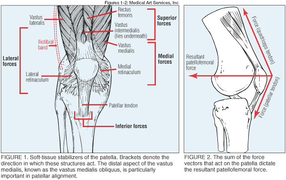
PFS often arises from a combination of intrinsic and extrinsic factors. Although quadriceps flexibility and function, genu varus, and hypermobile patellae have been identified by prospective studies as intrinsic risk factors for PFS,1,2 such studies are limited because of the disorder's multifactorial nature. Understanding PFS requires an understanding of the function of the patellofemoral joint (figure 1). It is important to appreciate the tethering effect of tendons and ligaments adjacent to the patella, as these are major determinants of forces across the joint (figure 2). In addition, overall lower-extremity alignment and the anatomic relationship between the trochlear groove and the posterior patella also produce variable forces across the patellofemoral joint. As the posterior aspect of the patella moves through the trochlear groove with knee movement, normal patellofemoral alignment and function disperse these forces across the articular surfaces.3
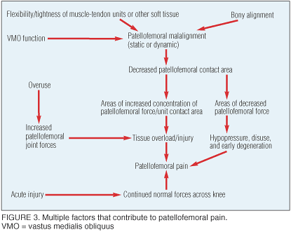
In patients with PFS, however, these static and dynamic factors most often produce increased forces across lateral joint surfaces and decreased forces across the medial side. Lateral pain may result from articular surface overload or adaptive shortening of the lateral retinaculum. Medial pain may result from articular surface hypoperfusion and early degeneration, or from chronic traction on the medial retinaculum.4,5 Although cartilage damage is frequently implicated in PFS, hyaline cartilage has no nerve endings, and therefore pain is generated by other structures, including the retinaculum, subchondral bone, synovium, or local small nerve endings.6 Several theories account for how biomechanics, overuse, and acute injury contribute to patellofemoral pain (figure 3).5,7
Assessing the Affected Patient
History. Pain in PFS is typically diffuse and poorly localized over the anterior aspect of the knee. Patients will sometimes report that the pain seems to be coming from "behind the kneecap." An early clue to a diagnosis is to ask the patient to point with one finger to the spot of maximal pain. Patients with PFS are often unable to pinpoint pain and instead will rub the front of their knees and complain of diffuse, rather than focal, pain within or over the anterior knee. Bilateral pain is common.
Patients with PFS will often report feeling knee swelling or "fullness," especially over the infrapatellar area, but significant swelling must raise questions of other intra-articular pathology. Buckling or subjective instability in PFS is often noted with stair use and is usually due to pain inhibition of the quadriceps. Patients will sometimes report "locking" or "catching" with PFS, which must be differentiated from the true mechanical block that can be seen with meniscal injury or loose intra-articular bodies.
Any changes in activity resulting in increased force across the patellofemoral joint should be identified. Such changes include altered training surface or terrain (especially hills and stairs), increased training volume, or other variations in training or daily activity (especially those leading to increased or prolonged knee flexion or squatting). However, in some patients a precipitating factor cannot be identified.
Patients should be asked about prior knee injuries. Previous patellar subluxation or dislocation or a fall directly onto the patella may indicate bone or articular cartilage damage that is exacerbated by normal patellofemoral forces.
Physical exam. Patients with anterior knee pain should have a complete physical examination of the knee, with particular attention to extensor mechanism function. The primary purpose of the physical exam is to look for possible sources of pathology and to assess for correctable factors that can be addressed with rehabilitation.
Assessing stance and gait. Stance and gait observations provide an overall impression of lower-limb alignment. Quadriceps bulk and symmetry; genu valgus, varus, or recurvatum; and increased foot pronation should be noted during this evaluation. Medial deviation of the knee during a one- or two-leg partial squat indicates weakness of the abductors and external rotators of the hip. Many patients with PFS will have pain with squatting, but this is also common with meniscal injury. Although much has been written about the significance of specific measures of patellofemoral alignment, several studies have shown little correlation between these measures and patellofemoral symptoms.8
Seated exam. Vastus medialis obliquus (VMO) bulk and patellar position should be assessed. Focused palpation of knee structures may reveal discrete areas of tenderness. Some causes of pain over the anterior aspect of the knee, such as pes anserine bursitis, medial plica syndrome, or Sinding-Larsen-Johansson disease, often produce focal tenderness. Fat pad and retinacular tenderness may produce tenderness over the anterior joint line.
Palpation over the patella during knee flexion and extension will often reveal crepitus, which is of debatable significance.6 One study9 that examined knee findings in normal subjects found that 40% of females had asymptomatic, and most often bilateral, patellofemoral crepitus. When crepitus is new, painful, or markedly asymmetric, however, it may signal other pathology.
Supine exams and tests. Physicians should assess active and passive range of motion of the knee and hip. Pain with hip testing, particularly with internal rotation, suggests primary hip pathology, which must always be considered in patients with knee pain. Knee ligaments and menisci should be thoroughly examined.
Flexibility assessment is an important, but often overlooked, component of the exam in patients with PFS. Particular attention should be paid to iliotibial band, hamstring, rectus femoris, and gastrocnemius and soleus flexibility. Tightness of these structures increases stress across the patellofemoral joint and is often readily treated with appropriate rehabilitation.
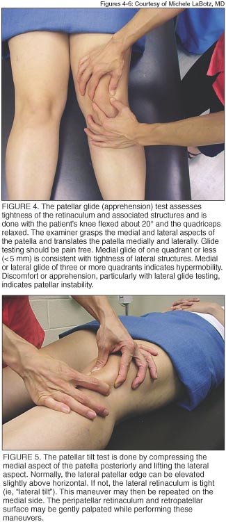
Specific tests of the patellofemoral joint may be performed. The patellar glide test (figure 4)10 and patellar tilt test (figure 5) assess movement of the patella. These tests primarily assess for retinacular tightness or laxity. The medial and lateral retinaculi are frequently tender and can be gently palpated using the same maneuver as the patellar tilt test. If the retinaculi are nontender, the posterior patellar facets can then be palpated as the patella is again tilted. Physicians should be aware that this retropatellar palpation is often uncomfortable, even in normal individuals, and examination of the contralateral knee can provide a baseline for patient tolerance to this test. Sagittal tilt can be assessed by observing distal patellar movement as the patient isometrically contracts the quadriceps. If the distal pole of the patella appears to move posteriorly, the action may be irritating the infrapatellar fat pad.
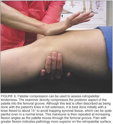
Compression testing done at differing degrees of flexion assesses pain as the patella moves through the femoral groove (figure 6).6 Since compression testing can be quite painful, it needs to be performed carefully and is often the final step in the physical examination.
Footwear. Physicians should also inspect shoes used for athletic activity and daily wear. The wear patterns are not often helpful in diagnosing PFS, but the condition and choice of footwear usually are. With athletic shoes, midsole cushion and support functions generally fatigue after 300 to 400 miles (about 3 months in the 30 mile/wk runner).
Imaging Studies
PFS is a clinical, rather than a radiographic, diagnosis. Most patients who have a typical history of overuse may be initially managed without imaging. However, if any red flags stand out in the history or exam—such as a significant mechanism of injury or effusion—physicians should undertake appropriate imaging studies to screen for other pathology. Physicians should be aware that abnormal patellofemoral alignment and articular cartilage changes, as assessed by radiographs and magnetic resonance imaging, are common in asymptomatic knees and do not appear to correlate with the clinical outcome after conservative treatment for PFS.11
Effective Treatment for Active Patients
Literature reviews of treatment for PFS reveal a paucity of high-quality trials on which to base treatment decisions.12-14 The relatively high rate of spontaneous recovery, especially in young patients with unilateral symptoms,11,13 and the multifactorial nature of PFS, confound therapeutic trials. Recent data suggest that patients with PFS do not have increased forces across the patellofemoral joint, but have an abnormal concentration of these forces onto a smaller articular surface area.7 This finding suggests that treatment should focus on dispersing joint forces across a larger surface area, and it supports the common treatment principle of addressing strength, flexibility, and other biomechanical factors that affect patellar tracking.
When formulating a treatment plan, the clinician needs to combine elements that have proven successful in controlled trials with information gathered from assessment of the individual patient. A key to successful treatment is to prepare patients to assume an active role in rehabilitation and to warn them that such a protocol may take several months to work. Although many patients with PFS will begin to note slight improvement shortly after initiating treatment, many studies of PFS therapies examine results after 6 to 12 weeks of nonoperative treatment.14,15 Several of these studies note continued improvement for up to 1 year.14,15
Elements of treatment. The first step in treatment is to reduce pain and inflammation. This includes regular and frequent application of ice for 15 to 20 minutes at a time, especially after activity. Nonsteroidal anti-inflammatory drugs (NSAIDs) or other analgesics often provide transient relief.
Patients should be encouraged to modulate activity by continuing pain-free activity and maintaining aerobic conditioning. Patients often get relief by decreasing training intensity and volume by about 50%. If pain persists, alternative activities should be sought. Patients who cannot tolerate continued activity and those with acute injury may require rest for up to 8 weeks, or until activities of daily living are pain free. Many runners with patellofemoral pain can tolerate freestyle swimming, bicycling (with the seat elevated to avoid excessive knee flexion), or elliptical trainers without discomfort. Stair-stepping machines and step aerobics should usually be avoided. Patients should also be counseled to avoid unnecessary force across the knee (eg, cross-leg sitting, prolonged squatting).
Home exercise programs should address strength and flexibility deficits and biomechanical factors (table 1) identified during the exam. This program can either be prescribed in the office (see the Patient Adviser, "Coping With Patellofemoral Syndrome") or with several visits to a physical therapist for instruction about a home exercise program. Whichever approach, the program should include the following:
- Restoring quadriceps strength. This action is a key component to reducing symptoms and improving long-term outcome in PFS.11 Although straight-leg raises and other open-chain exercises targeting the VMO are commonly used in PFS rehabilitation, patients may do better with closed-chain exercises such as partial squats or step-downs.16
- Strengthening external hip rotators. Athletes who have weak external hip rotators often benefit from appropriate strengthening exercises.6
- Stretching tight regions. Problem structures often include the iliotibial tract, quadriceps, hamstrings, gastrocnemius-soleus complex, and lateral retinaculum.17