A Patient's Guide to Plantar Fasciitis (Heel Pain)
Introduction
Plantar fasciitis is a painful condition affecting
the bottom of the foot. It is a common cause of heel pain
and is sometimes called a heel spur. Plantar
fasciitis can come from a number of underlying causes.
Finding the precise reason for the heel pain is sometimes
difficult. Even so, several options are available for
treatment.
This guide will help you understand
- how plantar fasciitis develops
- how the condition causes problems
- what can be done for your pain
Anatomy
Where is the plantar fascia, and what does it do?
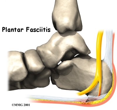
The plantar fascia is a structure that runs from
the front of the heelbone (calcaneus) to the ball of
the foot. This dense strip of tissue helps support the arch
of the foot by acting something like the string on an
archer's bow.
As you can imagine, when the foot is on the ground a
tremendous amount of force (the full weight of the body) is
concentrated on the plantar fascia. This force stretches the
plantar fascia as the arch of the foot tries to flatten from
the weight of your body. This is just like the string on a
bow is stretched by the force of the bow trying to
straighten. This leads to stress on the plantar fascia where
it attaches to the heelbone. Small tears of the fascia can
result. These tears are normally repaired by the body.
As this process of injury and repair repeats itself over
and over again, a bone spur (a pointed outgrowth of the bone)
sometimes forms as the body's response to try to firmly
attach the fascia to the heelbone. This appears on an X-ray
of the foot as a heel spur.
Related Document: A
Patient's Guide to Foot Anatomy
Causes
How does plantar fasciitis develop?
Heel pain probably comes from several causes. In some
cases the heel spur can be so big it causes pain itself, but
this is rare. The chronic inflammation of the fascia itself
may be the source of pain in many cases. (This condition is
probably most accurately called plantar fasciitis.)
As we age, the very important fat pad that makes up the fleshy portion of the heel
becomes thinner and degenerates. This can lead to inadequate
padding on the heel and chronic pain in this area.
Some physicians feel that the small nerves that travel
under the plantar fascia on their way to the forefoot become
irritated and may contribute to the pain. In many cases, the
actual source of the painful heel will never be clearly
defined without doubt.
Symptoms
What does plantar fasciitis feel like?
The symptoms of plantar fasciitis include pain in the
center of the heel when weight is placed on the foot. This
is usually most pronounced in the morning when the foot is
first placed on the floor.
Diagnosis
How do doctors diagnose the condition?
The diagnosis of plantar fasciitis is generally made
during the history and physical examination. There are
several conditions that can cause heel pain, and plantar
fasciitis must be distinguished from these conditions.
An X-ray may be ordered to rule out a stress fracture of
the heel bone and to see if a bone spur is present that is large enough to cause
problems. Laboratory investigation may be necessary in some
cases to rule out a systemic illness causing the heel
pain, such as rheumatoid arthritis, Reiter's syndrome, or
ankylosing spondylitis. These are diseases that affect the
entire body but may show up at first as pain in the
heel.
Treatment
What can be done for my pain?
Nonsurgical Treatment
Most patients get better with the help of nonsurgical
treatments. Stretches for the calf muscles on the back of
the lower leg take tension off the plantar fascia.
A night splint can be worn while you sleep. The
night splint keeps your foot from bending downward, and it
places a mild stretch on the calf muscles and the plantar
fascia. People seem to get better more quickly when using a
night splint, and they report having less heel pain when
placing their sore foot on the ground in the morning.
Supporting the arch with a well fitted arch support, or
orthotic, may also help reduce pressure on the
plantar fascia. Also, placing a special type of insert into
the shoe, called a heel cup, can reduce the pressure
on the sore area and add padding to a heel that has lost
some of the fat pad through degeneration.
Shock wave therapy is a newer form of nonsurgical
treatment. It uses a machine to generate shock wave pulses
to the sore area. Patients generally receive the treatment
once each week for up to three weeks. It is not known
exactly why it works for plantar fasciitis, but recent
studies indicate that this form of treatment can help ease
pain, while improving range of motion and function.
Anti-inflammatory medications are sometimes used to
decrease the inflammation in the fascia and reduce your
pain. An injection of cortisone into the area of the fascia
is effective. Cortisone should be used sparingly since it
may contribute to the process of degeneration of the fat
pad, actually making the problem worse.
Surgery
Surgery is a last resort in the treatment of heel pain.
Physicians have developed many procedures in the last 100
years to try to cure heel pain. Most procedures that are
commonly used today focus on several areas:
- remove the bone spur (if one is present)
- release the plantar fascia
- release pressure on the small nerves in the area
Usually the procedure is done through a small incision on
the inside edge of the foot, although some surgeons now
perform this type of surgery using an endoscope. An
endoscope is a small TV camera that can be inserted into a
joint or under the skin to allow the surgeon to see the
structures involved in the surgery. By using the endoscope,
a surgeon can complete the surgery with a smaller incision
and presumably less damage to normal tissues. It is unclear
whether an endoscopic procedure for this condition is better
than the traditional small incision.
Surgery usually involves identifying the area where the
plantar fascia attaches to the heel and releasing the fascia partially from the bone. If a small spur is present this is removed. The small
nerves that travel under the plantar fascia are identified
and released from anything that seems to be causing pressure
on the nerves. This surgery can usually be done on an
outpatient basis, meaning you can leave the hospital the
same day.
Rehabilitation
What should I expect after treatment?
Nonsurgical Rehabilitation
Patients with plantar fasciitis are commonly prescribed
physical therapy. Therapists design exercises to improve
flexibility in the calf muscles and the plantar fascia.
Treatments directed to the painful area help control pain
and swelling. Examples include ultrasound, ice packs, and
soft-tissue massage. Therapy sessions sometimes include
iontophoresis, which uses a mild electrical current
to push anti-inflammatory medicine to the sore area.
A customized foot orthotic may be designed to support the
arch of the foot and to help cushion the heel. Or your
therapist may recommend you use a heel cup.
Ideas are offered for you to use at home, such as doing
your stretches for the calf muscles and the plantar fascia.
You may also be fit with a night splint to wear while you
sleep. As mentioned earlier, the night splint is designed to
put a gentle stretch on the calf muscles and plantar fascia
as you sleep.
After Surgery
It will take several weeks before the tissues are well
healed. The incision is protected with a bandage or dressing
for about one week after surgery. You will probably use
crutches briefly, and a physical therapist may be consulted
to help you learn to use your crutches.
The stitches are generally removed in 10 to 14 days.
However, if your surgeon used sutures that dissolve, you
won't need to have the stitches taken out. You should be
released to full activity in about six weeks. | 

)
)
)
)
)