PMC full text:
Published online 2020 May 31. doi: 10.4103/jfmpc.jfmpc_95_20
Figure 2
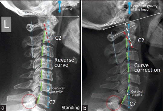
Cervical spine lateral view of Case 2. (a) Prior to treatment lateral radiograph displayed a reverse cervical lordosis, degenerative spondylosis with ankylosis of the C7/T1 facet joints (red circle). The cervical gravity line (blue dotted line) just touched the anterior body of the C7. (b) The repeat lateral radiographs 9 months later exhibited improved general cervical lordosis. A smooth vertical alignment of each posterior body corner was noted. The cervical gravity line fell within the C7 vertebra. Redlund-Johnell criterion (white dotted line) was reduced by 6.55% and Ranawat index (red dotted line) was reduced by 8.88%