PMC full text:
Published online 2020 May 31. doi: 10.4103/jfmpc.jfmpc_95_20
Figure 3
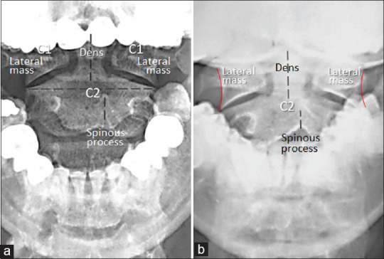
Open mouth radiographs of Case 2 (a) and of Case 3 (b) at initial presentation. There was symmetrical spacing of lateral zygapophyseal (C1-C2) joints and of odontoid-lateral mass intervals. Note a deviation of the C2 spinous process with respect to the alignment of the dens, a suggestive finding of C2 rotation