PMC full text:
Published online 2020 May 31. doi: 10.4103/jfmpc.jfmpc_95_20
Figure 4
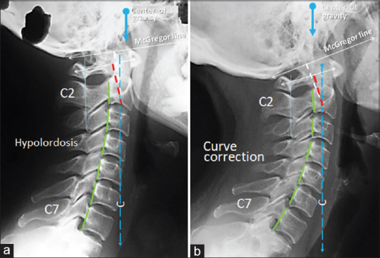
Initial and follow-up radiographs of Case 3. (a) Initial radiograph showed a loss of cervical lordosis, osteophytic lipping of the vertebrae, narrowing of the joint space of the C7/T1, and facetitis of the right C5/C6 and C7/T1 facet joints. (b) The repeat radiography 2 years later exhibited improved general cervical lordosis. There was a smooth vertical alignment of each posterior vertebral corner. Redlund-Johnell criterion (white dotted line) was reduced by 6.34% and Ranawat index (red dotted line) was reduced by 10.41% FHP (Forward head posture)