PMC full text:
Published online 2024 Apr 8.
Figure 9
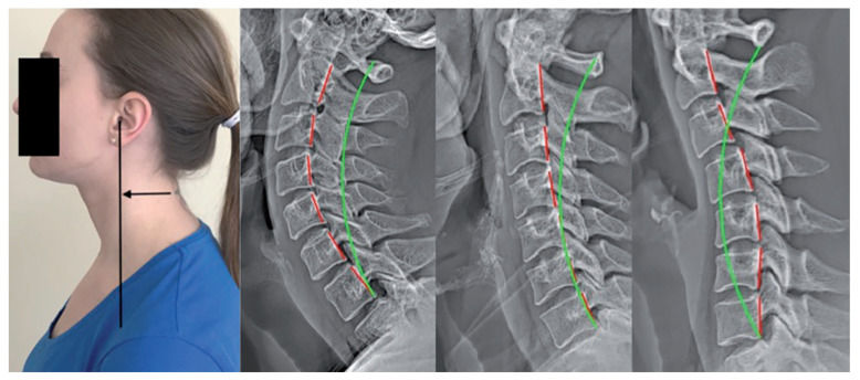
Forward head posture as shown in a posture photograph and three unique lateral cervical radiographs. All three X-ray images have about 25 mm of forward head translation, as measured with the C2–C7 SVA. Left radiograph: hyperlordosis; middle radiograph: hypolordosis; and right radiograph: kyphosis. The green line is a standard alignment; the red line highlights the patient’s alignment along the posterior body margins of cervical vertebra C2 through C7.