| PMC full text: | Published online 2009 Apr 22. doi: 10.1128/JVI.00475-09
|
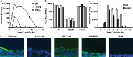
FIG. 1.
HPIV3 wt and HN variant virus growth in HAE and cotton rat models.
(a) HAE cultures were infected with wt HPIV3 or with the HN T193A or H552Q variant. At the indicated days postinfection (x axis), the number of infectious particles released (y axis) was determined by plaque assay. Data points are means (± standard deviations) of triplicate measurements and are representative of at least five experiments.
(b) Cotton rats were infected with wt HPIV3 or with the HN T193A or H552Q variant, and at 3 days postinfection, the viral titer (PFU/g lung tissue) (y axis) was determined by plaque assay. Each bar represents one animal.
(c) HAE cells were infected with the HN N551D variant or wt HPIV3. At the indicated time points after infection (x axis), the number of infectious particles released (y axis) was determined by plaque assay. Data points are means (± standard deviations) of triplicate measurements and are representative of data from three experiments.
(d) HAE cultures infected as described above (a and c) were fixed at day 5 postinfection and processed for immunofluorescent detection of viral antigens. Viral antigens were visualized using FITC (green), and nuclei were counterstained with DAPI (blue).
