Figure 1
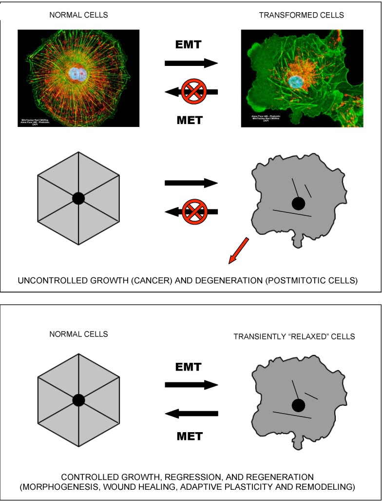
Epithelial-to-mesenchymal-like organizational transitions
The shown schematics and cell images illustrate the concept of EMT-like, organizational transitions, which involve coordinated switches in cell adhesions, cytoskeletal organization/dynamics, metabolism, and signaling. The cell image shown on the left is a normal kidney fibroblast in cell culture (CV-1 cells), whereas the cell image on the right is its SV40-transformed derivative (COS-7 cells).
Note the dramatic structural collapse of the mitochondrial and actin filamentous networks in transformed cells
(F-actin – GREEN;
mitochondria – RED).
Images courtesy of Carl Zeiss Microscopy GmbH.