Figure 3
Integration of keratin cytoskeletal networks in cultured cells
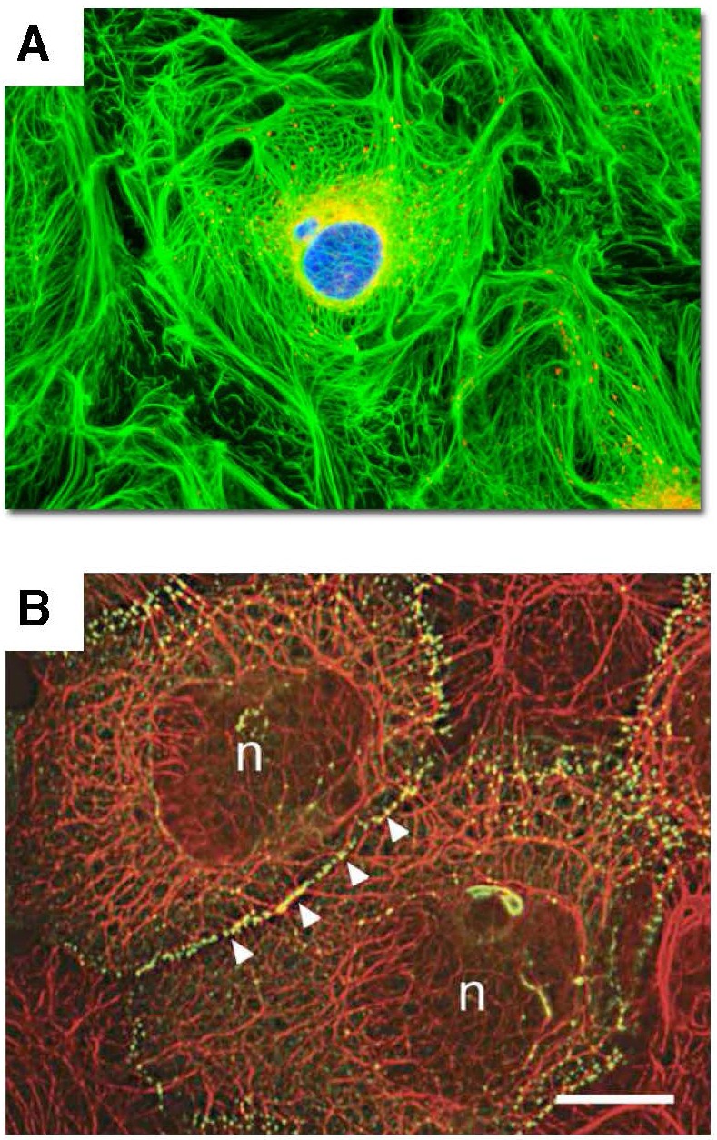
Cell images illustrate the integration of individual intermediate filament networks via dynamic cell adhesions into large-scale, collective, cytoskeletal architectures. A.
Rat kangaroo kidney epithelial cells in culture.
Keratin – GREEN;
mitochondria – RED.
Image courtesy of Olympus America; B.
Skin epidermal keratinocytes in culture.
Keratin – RED;
cell adhesions – GREEN;
(n) nucleus.
Reproduced from Kim S. and Coulombe P.A. [81].
Courtesy of Dr. Kathleen Green.