| PMC full text: | Cell Death Differ. Author manuscript; available in PMC 2009 Oct 5. Published in final edited form as: Cell Death Differ. 2009 May; 16(5): 655–663. Published online 2009 Jan 16. doi: 10.1038/cdd.2008.191 |
Figure 1
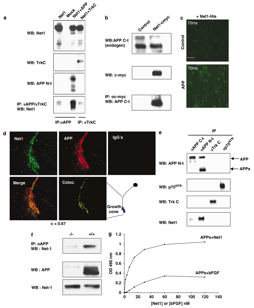
Netrin-1 interacts with APP.
HEK293T cells were transiently transfected with myc-tagged netrin-1 and/or APP and/or TrkC. Cell lysate was utilized for immunoprecipitation, using either an anti-N-terminal APP antibody or an anti-TrkC antibody. Immunoblots were probed with antibodies raised against N-terminal APP, netrin-1, or TrkC.
(b) HEK293T cells were transfected with netrin-1-expressing construct or not, and endogenous APP was, after c-myc (netrin-1) pull-down, revealed using an anti-C-terminal APP antibody.
(c) B103 cells were transfected with mock- or APP-expressing plasmid and treated for 1 h with his-netrin-1. Anti-HIS immunohistochemistry is shown.
(d) Colocalization of 5A3/1G7 (APP extracellular domain) with netrin-1 in growth cones of primary cortical neurons. Primary cultures of neurons from DBA/2J embryos were fixed in 4% PFA and stained with 5A3/1G7 and antinetrin 64 or with mouse and rabbit IgGs followed by Alexa568- and Alexa488-conjugated antimouse and antirabbit secondary antibodies, respectively. Stacks of images (z-step=0.25 µm) were acquired with a laser scanning confocal microscope. Analysis of colocalization was performed using the Coloc algorithm in Imaris Bitplane. The Pearson correlation coefficient of channels A (green) and B (red) inside the colocalized region was used as a measure of the degree of colocalization.36 Panels shown are Net1 (netrin-1), APP, merge, Coloc. (colocalization channel), and IgGs (mouse and rabbit IgGs). A cartoon of the region of a cortical neuron shown in the various panels is indicated.
(e) Cortexes from E16.5 mouse embryos were collected and semidissociated, and cells lysates were submitted to immunoprecipitation using anti-APP (C-terminal or N-terminal), anti-TrkC or anti-p75NTR antibody for the pull-down. Immunoblot were then performed using either APP, p75ntr or TrkC antibody.
(f) Immunoprecipitations were performed with a mixed mouse monoclonal anti-APP (5A3/1G7) antibody from E16.5 mouse brain of either wild-type embryo (+/+) and APP mutant (−/−). Netrin-1 and APP immunoblot using a monoclonal antinetrin-1 (dilution 1/1000) (R&D system) and monoclonal anti-APP (5A3/1G7) antibodies are shown.
(g) An Elisa assay was developed to determine the KdAPP/netrin. In all, 2.5 µg/ml of αAPPs protein was coated in 96-wells plate and various netrin-1 concentrations were added. Similar experiment was performed using the pair APP/bFGF. Quantification of the interaction is indicated here by the measurement of the optic density (intensity). Determination of Kd was derived from a simulated Scatchard plot (bound/estimated free = f(bound))