| PMC full text: |
|
Figure 4.
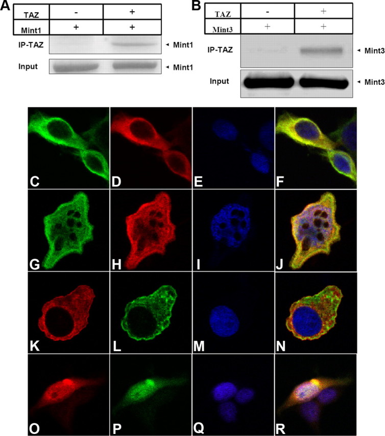
TAZ and YAP interact with Mint1 and Mint3. A, B, Coimmunoprecipitation of TAZ with Mint1 (A) and Mint3 (B). 293T cells transiently expressing full-length FLAG-tagged TAZ and Mint1 (A) or Mint3 (B) were lysed and the crude lysates were subjected to immunoprecipitation with M2 anti-FLAG beads. Cells expressing Mint1 (A) or Mint3 (B) only were used as controls. Immunoprecipitated samples were subjected to SDS-PAGE and analyzed by immunoblotting with anti-Mint1 (A) or anti-Mint3 (B) antibodies. Total cell lysates were used as input controls. C–F, TAZ colocalizes with Mint1. Double labeling of 293T cells cotransfected with Mint1 (green) and TAZ (red) constructs. G–J, TAZ colocalizes with Mint3. Double labeling of 293T cells cotransfected with Mint3 (green) and TAZ (red) constructs. K–N, YAP colocalizes with Mint1. Double labeling of 293T cells cotransfected with Mint1 (red) and YAP (green) constructs. O–R, YAP colocalizes with Mint3. Double labeling of 293T cells cotransfected with Mint3 (red) and YAP (green) constructs. DAPI staining (blue) was used to visualize nuclei. Images were captured on a laser-scanning confocal microscope (Zeiss LSM510).