

Recognition of Perinatal Stroke in a Chiropractic Practice:
Case Report and Clinical Challenges Related to a Late DiagnosisThis section is compiled by Frank M. Painter, D.C.
Send all comments or additions to: Frankp@chiro.org


FROM: J Clinical Chiropractic Pediatrics 2012 (Jun); 13 (1): 958–967 ~ FULL TEXT
OPEN ACCESS Lise Hestbaek, PhD, Annette Jřrgensen, DC, and Jan Hartvigsen, PhD
Nordic Institute of Chiropractic and Clinical Biomechanics,
Odense, Denmark.
Introduction: In recent years, improvements in medical techniques and technology have enabled primary health care practitioners to diagnose perinatal strokes in infants far earlier than ever before. This new technology can also support chiropractors, especially those working with pediatric patients, in order to validate their diagnosis when they recognize these initial symptoms.
Objective: The aim of this paper is to raise awareness and assist doctors of chiropractic in recognizing the physical and behavioral signs of perinatal stroke as they present in a chiropractic office for assessment and treatment. The paper will relate the case of a 7–month-old infant who, after visiting a chiropractic office with apparent hemiparesis and delayed developmental milestones, was post-medically diagnosed as having suffered a presumed perinatal stroke.
Discussion: Early recognition of perinatal stroke is vital since late diagnosis can lead to a lifetime of debilitating neurological conditions as well as potential increased direct and indirect costs to society. For those who receive a late diagnosis, a chiropractor, as a member of a multidisciplinary team, can play a major role in rehabilitation by supporting the function of the nervous system, reducing muscle and joint rigidity and optimizing neuronal plasticity.
There are more articles like this @ our
STROKE AND CHIROPRACTIC PageConclusion: It would be prudent for chiropractors to receive more training in the detection of perinatal stroke. Early detection can be accomplished by way of a comprehensive questionnaire and focused physical examination. The early detection of possible perinatal stroke will permit a referral to the proper professional and rapid intervention which will increase the likelihood of a positive outcome. More studies need to be done to reach a better understanding of the pathophysiology of perinatal strokes. Clinical guidelines also need to be established that will improve the prognosis for pediatric patients.
Key words: perinatal stroke, infantile stroke, fetal stroke, presumed prenatal stroke, chiropractic, west syndrome, hemiparesis, cerebral palsy, delayed milestones, rehabilitation
From the FULL TEXT Article
INTRODUCTION
Perinatal strokes are now estimated to occur in 1 in 4,000–5,000 newborn babies. [1–4] As a result of today’s new imaging technologies, the rate of diagnosis of this condition is on the rise. Because symptoms may not appear until a baby is between the ages of 4 to 6 months, a chiropractor may be the first health care professional that parents seek out when they encounter difficulties while breastfeeding or become aware of a delay in reaching expected developmental milestones. Chiropractic is a health care profession that focuses on disorders of the musculoskeletal system and the nervous system, and the effects of these disorders on general health.
At a perinatal stroke workshop held in 2007, perinatal ischemic stroke was defined as a group of heterogeneous conditions where neither a focal disruption of cerebral blood flow secondary to arterial ischemic stroke (AIS) or cerebral venous thrombosis (CVT) occurs nor embolization (hemorrhage) between 20 weeks of fetal life to the 28th post-natal day, and confirmed by neuroimaging or neuropathological studies. [5–7] If blood flow is interrupted for a period of time longer than a few seconds, the brain can be permanently damaged. Early recognition is important and contributes to an improved prognosis. 80% of all perinatal strokes are ischemic while the remainder are hemorrhagic or venous. [8]
As recommended by Raju et al. (2007), perinatal strokes can be classified more effectively by the timing of their detection rather than by the time of onset. [5] Therefore, we divide them into three categories; fetal stroke, neonatal stroke and presumed fetal or neonatal stroke. The last category involves obtaining evidence from an imaging study of a long-standing stroke without previous clinical symptoms. In these cases, though, suspicions were raised because of the higher prevalence of chronic neurological deficits and cerebral palsy. [5, 7, 9]
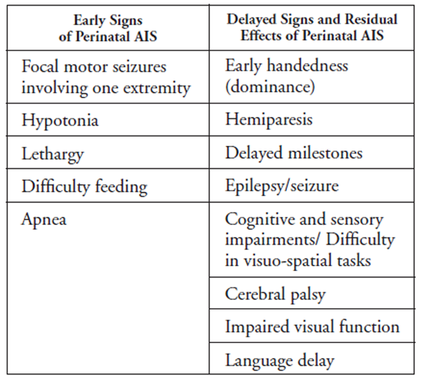
The clinical presentation of a perinatal stroke depends on the age of the child at the time of diagnosis. In newborns that are diagnosed in the early stages, 25 to 40% have seizures. [3] This is the most common trigger for the provision of more detailed assessment. Most seizures are focal and infants may appear well between seizures. Other systemic signs, if present, are nonspecific and subtle and may include difficulty feeding, hypotonia, lethargy, or apnea. In neonatal strokes diagnosed retrospectively (also called presumed neonatal stroke), the presentation includes asymmetry of reach and grasp, hemiparesis, failure to reach developmental milestones, or post-neonatal seizures. [3]
Medical management consists of aggressively treating the symptoms, mostly seizures. In the presence of refractory hemispheric epilepsy, hemispherotomy and functional hemispherectomy could be indicated to reduce or eliminate seizures and promote neurological development [10] in the infant, and later, the child. To our knowledge, the current literature contains no studies or case reports on chiropractic management (CINAHL (EBSCO), Medline, Pubmed, Index to chiropractic literature). It is possible that chiropractors can play a major role in increasing brain plasticity thereby optimizing long term functionality as part of a multi-disciplinary team approach. Today’s chiropractors need to remain current on the information pertaining to this condition in order to assess patients utilizing detailed case history, a thorough physical examination to clarify the diagnosis, recommend referrals, and be members of the rehabilitation team alongside other health care professionals.
For the purposes of this paper, childhood stroke will not be discussed; detailed information is provided elsewhere. [1, 8, 11]
Case Presentation
A female infant was born at 38 weeks, 4 days gestation to a 37–year-old mother, gravida 4 para 4. The history of the pregnancy was unremarkable with the exception of some medication administered for nausea (Diclectin®) which was taken in the first trimester. Two ultrasounds were performed at 12 and 28 weeks respectively and were normal. The birth was a normal vaginal delivery with APGAR scores of 9–10 after 1 and 5 minutes. Total labor time was 6 hours with 45 minutes of pushing; the baby presented posteriorly and had to be assisted by manual extraction. No medication/ sedation was administered during labor, no stimulation of labor or epidural anesthesia; no placental examination was performed. The mother was discharged from the hospital within 72 hours, without any apparent problems. Breastfeeding was difficult at first. When the baby initially latched, the suckle was weak attributed to the sleepy state of the baby, but the mother persevered and was ultimately successful. In this case, the mother was experienced, knowledgeable and was a breastfeeding support mother in a lactation group. During her hospital stay, she was expressing milk and giving it to the infant with a spoon. In her first month of life, the baby also required an intervention for a posterior tongue tie which also contributed to difficulties with latch and transfer of milk.
In the first months, the mother reported some slight delays in the baby’s attainment of expected developmental milestones. She noticed that her baby was not as alert as her other children were at the same age, but she was able to smile and interact with others. By comparison, eventually she noticed that some significant milestones were delayed, these included; head holding, an absence of ventral positioning and an asymmetry in the use of her upper extremities. She reported that a few weeks after birth, the baby was not using her right arm as much as the left one and that her hand was mostly held in a fist and maintained in flexion. At the age of six months, the baby was moving her arm at the elbow joint and her hand was able to open more frequently. During her last medical visit at 6˝ months, the medical doctor expressed no concern about the infant’s condition and the delayed milestones and suggested a follow up a month later. She demonstrated no ability to sit and was not comfortable lying in the prone position (“tummy time”). It is notable that the child was the family’s 4th daughter and that the mother, based on her previous experiences, was aware that something was wrong.
The baby was brought to our office for a physical evaluation at 7 months. Observations showed reduced facial expressions with an asymmetry of the eye gaze, the right eye having a tendency to diverge at some point and a delay in the active cervical motion when following objects. She was able to smile although she was reluctant, not often making eye contact. The level of activity of the upper extremity was reduced and asymmetrical although she was using the upper extremity mostly at the elbow and within 60 degrees of shoulder flexion on the right side. The baby was uncomfortable in the ventral position and displayed a lack of extension at the occipital and cervicothoracic areas. She wasn’t able to turn on either side and failed to attempt to do so even when stimulated. The infant could not sit up, with or without support, and she was unable to be pulled up by hand into a sitting position. She was able to bring objects close to her mouth with her left hand only.
The cranial evaluation showed normal fontanels. Some cranial restrictions were noted in the following bones; parietal, frontal, temporal, occiput, and sphenoid. Light palpation of the cranials elicited a withdrawal reflex accompanied by crying and palpation of the upper cervical region as well. Tongue movement was within normal limits and sucking occurred during breastfeeding though the infant refused to do so when stimulated digitally.
The range of motion of the cervical spine was restricted in right rotation and in flexion/extension with tension at the suboccipital area. The occiput was slightly fixed in extension. The range of motion (ROM) of the right shoulder, clavicle and scapula were restricted and elicited a crying response from the baby. The pectoralis, SCM, scalene, subclavian, biceps, subscapularis, upper trapezius, and levator scapula muscles were all hypertonic on the right side; The ROM of the right hip was also reduced mostly in long axis traction and internal rotation but both legs were restricted and positioned in abduction and external rotation.
During the neurological examination, the Babinski reflex was present bilaterally but slightly reduced on the right side. The palmar grasp reflex, the Moro reflex and the ATNR (asymmetric tonic neck reflex) were reduced on the right side. Deep tendon reflexes were not performed at the initial examination.
The chiropractic examination showed vertebral subluxations upon palpation at C0–C2, C6–T1, T3–5, T9–T10 with a posterior left sacrum at the S1 segment.
The first working diagnosis at that time was of an obstetrical brachial plexus injury with biomechanical dyskinesia (vertebral subluxation complex) around the C0– C2 and C7–T5 areas also involving the shoulder area complex. Other differential diagnosis included complications of consolidated clavicle fracture, a possible cerebral lesion like cerebral palsy or tumor. Two weeks after the first visit, some additionally symptoms were observed and relayed by the mother. The mother noticed that her baby was having some absences during the day followed by some flexor spasms of the neck, mostly when she was tired or upon awakening. The possibility of a cerebral lesion then became the first probable diagnosis. With videos of the spasms of hand, she was then able to see a pediatric neurologist who ordered scans. At 7˝ months, the baby girl was diagnosed with West syndrome and spastic hemiplegia caused by a presumed fetal stroke. The ischemic area involved was in the territory of the left middle cerebral artery and the majority of her left parietal lobe was liquefied by the time of diagnosis.
Interventions and outcomes
After the initial examination of the baby, we made it clear to the mother that co-management of medical and chiropractic professionals was necessary in order to clarify the diagnosis and to gather more facts on the extent of the condition. Fortunately, within three weeks, we received the medical diagnosis and the multidisciplinary approach became essential, optimizing a long-term functional outcome and impacting the brain and nervous system plasticity. In view of the fact that the condition is non-reversible, the management of the baby’s condition would consist of follow-ups on a long term basis. Rehabilitation would potentially improve the deficiencies and help the baby develop to the maximum of her potential while simultaneously improving the quality of her life. Given the state of the health care system (Quebec), delays in multidisciplinary referrals are common. Consequently, the chiropractor was the only health care provider working with the patient from the age of 7˝ months to 9˝ months at which time she was seen by the consulting physician and the multidisciplinary management began.
Chiropractic management
Initially, we saw the patient twice a week for 6 weeks, once a week for 5 months and then every two weeks for 6 months. Between the ages of 18 months to 28 months, we saw her for adjustments at intervals of 3 to 5 weeks working around the mother’s and child’s many other associated appointments and interventions.
Chiropractic management started gradually with the application of the following techniques:
Fascial release to upper and lower extremities; slight mobilization on clavicle, arm, scapula, elbow, wrist and hip joints
Slight traction of occiput-sacrum in the cephalic direction
Chiropractic adjustments in the thoracic and sacral regions T2–3, T8–T10 and sacrum (low force manual techniques)
C1 and C6–7: Activator method in cervical spine or sustained pressure especially C1 on right side in laterality and C6–7
Low force manual cranial techniques
------------------------------------------------------
** Note: Neither manual nor activator adjustments were delivered to correct the atlas laterality in the first 2 weeks; we decided to proceed with additional cranial work after the diagnosis of a possible fetal stroke while waiting for information on the probable cause(s).
------------------------------------------------------
After the initial 6 weeks we added the following:
Joint mobilizations exercises (mostly arm and hip)
Stimulation exercises to stimulate right and left hemispheric communication and the vestibular system performed in the clinic and given to the mother to do at home:
Frog and spider exercises
Contralateral hand to toe touch
Hip and arm passive mobilizations
Ventral exercises on a roll + gym ball
After the first two weeks of working with the infant, the mother immediately noticed improvement in shoulder mobility through active motion. By helping her with passive stretching and movements, the mother noticed less resistance. Of note, during the first visits, the baby cried simply after being touched or as a result of movement in the shoulder-clavicle-scapula area. This intense crying decreased in the first two weeks. The ROM in the cervical area improved rapidly with less resistance than was present at the outset of the treatment. No adverse effect from any of the treatments provided was reported by the mother.
Two thermal scans were performe (Millenium Insight®) at the ages of 12 and 15 months. Both scans showed hyperactivity of the autonomic nervous system in the upper cervical and the cervicothoracic regions.
Medical management
Co-management was recommended soon after the initial examination in order to further clarify the diagnosis. During the third week of care, the mother saw a neurologist about the cervical flexion spasms that had now begun: the head was falling into flexion and left lateral flexion with a superior eye-glaze in a cluster-like pattern, mostly when she was waking up. At 7˝ month, a brain EEG confirmed hypsarrythmia patterns typical of infantile spasms\West syndrome related to a possible intrauterine or fetal stroke. Hypsarrythmia is an abnormal interictal pattern, consisting of high amplitude and irregular waves and spikes in a background of chaotic and disorganized activity seen on electroencephalogram (EEG), frequently encountered in an infant diagnosed with infantile spasms. Blood tests were done at 8 months to confirm a lowered resistance to the C-protein. A cerebral MRI was done at 9˝ months and it confirmed a left middle cerebral artery (MCA) lesion that affected most of the parietal lobe. Involvement of the factor V Leiden (FVL) was confirmed at the time for the baby and the mother also tested positive for the Leiden V factor a few months later. Maternal and neonatal thrombophilia in the presence of FVL has been investigated scientifically in the pathogenesis of perinatal arterial ischemic stroke.
Initial management consisted of controlling the spasms with pharmacological agents. A trial of three different types of medication was run:
Sabril® (Vigabatrin) was first introduced;
Lamictal® (Lamitrogine) as a secondary medication;
The spasms were difficult to control so a third medication was prescribed for epileptic spasms at 12 months: Keppra. XR® (Levetiracetam)
Keppra® was removed after a trial of a few days because the baby was exhibiting side effects (anorexia, insomnia, sudden mood changes and unusual behavior). At that time, the neurologist recommended that the baby undergo surgery to control the epilepsy that was preventing the brain from developing. A functional hemispherectomy was performed on the child at 15˝ months. The first two medications were administered for a year following surgery and were gradually eliminated; Sabril® at six months postsurgery and Lamictal® at twelve months.
Multidisciplinary Approach
In order for the baby to reach maximal function, a multi-disciplinary approach was essential. Since the initial application of chiropractic adjustments and recommendations, several health care professionals were added:
Physical therapist (conventional and water rehabilitation)
Occupational therapist
Speech therapist
Opthalmologist
Neuropsychologist
Audiologist
At 13˝ months, right before surgery, the infants expressive language included the words “mommy and “yes” and nodded her head yes and no. But the seizure activity was so frequent that it was preventing the brain creating and maintaining developing neuronal connections. Subsequent to surgery, motor and developmental improvements were noted. The baby was relating to people more readily; she was more alert, started to babble more and developed both her receptive and expressive language skills. At 20 months she started sitting unattended. At 27 months, she is still unable to crawl, kneel or roll.
Discussion

Table 1 Perinatal arterial ischemic stroke (AIS) is defined as a fetal or neonatal cerebrovascular event and is 17 times more common than strokes later in childhood and represents 80% of neonatal strokes [1–3] with a recurrence rate of 3–5% in later childhood. Some cases present in the first days of life but many can be delayed in their presentation. Hand preference will assist in establishing a diagnosis of hemiparesis. To complicate matters, in some cases there are no episodes of seizure as in the clinical presentation of our case. Infants presenting neurological signs and seizures (or epilepsy) soon after birth can be diagnosed as early as their stay in the hospital. Because delayed presentations can be revealed first in our chiropractic practices, it is essential that chiropractors be trained to recognize the associated signs and symptoms of AIS (including seizures). Parents typically come to us after having noticed delayed milestones or other early physical or behavioral concerns. The signs and symptoms of AIS are outlined in Table 1. [3, 9]
Cognitive impairment after a neonatal stroke ranges from 0 to 55% and language delay up to 25%. More than 50% of cortical strokes are located in the middle cerebral artery (MCA) territory with complete and posterior truncal area being more common. [7] Children who suffer a neonatal stroke or infarction in the MCA territory may develop thalamic atrophy but whether this has long-term implications for sensory integration is not clear and difficult to assess in this age group. The incidence of cerebral palsy after perinatal AIS ranges widely, from 6 to 88% according to the literature. Most infants will walk by the age of 2 and will eventually be able to be assimilated into a regular classroom. Only a small percentage will have to undergo surgery for major and intractable epileptic syndromes like West syndrome as in our case study. [12] A study done on 40 children who had suffered perinatal stroke found that the extent of the stroke and injury to any of the a number of regions (Broca’s region, the internal capsule, Wernicke’s area or basal ganglia) were associated with cerebral palsy. [13] Children who appear normal in the neonatal period but develop a hand preference or have a seizure after 2 months of age as a result of perinatal AIS may have a worse prognosis than children who displayed neurological signs as neonates. In such cases, the presenting hemiparesis is more likely to persist. [13, 14] This was also the situation in our case-report.
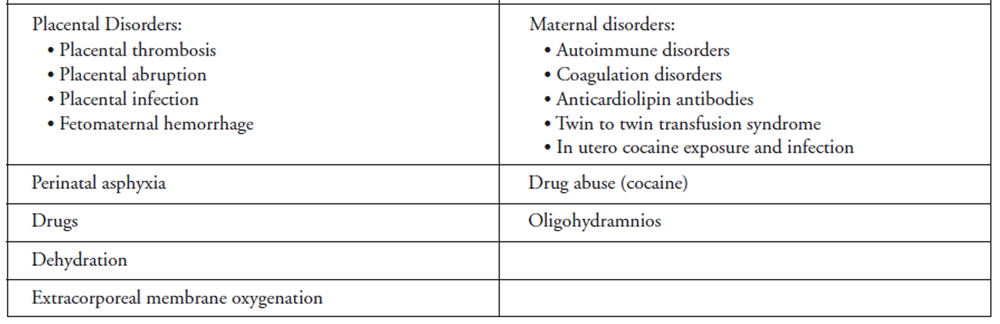
Table 2A
Table 2B The causes of perinatal AIS are presently poorly understood. Risk factors for perinatal strokes can be classified under maternal conditions, complications during pregnancy and delivery and fetal conditions as shown in Table 2. [1, 3, 4] It must also be noted that multiple risk factors increase the rate of AIS.
Infantile spasms/West syndrome
West syndrome is described by a characteristic triad of infantile spasms, interruption in psychomotor development and hypsarrythmias. Only 5 to 12% of patients exhibit normal mental and motor development, 50% are left with motor impairment and 70 to 78% are mentally challenged. [15] The severe spasms related to this condition are usually resistant to usual seizure medication and the prognosis is poor and associated with increased morbidity because maturation and cognitive development of the brain are affected by the spasms. This category of epileptic encephalopathy responds more favorably to hormonal treatment like ACTH and steroids than conventional antiepileptic medication. [16] However, for some geographic and availability reasons, our case study patient was prescribed Vigabatrin, and ACTH/steroids were never considered by the medical team unlike in many other cases described in the literature. [15–17] Most cases respond well to medication but in some cases, the patients have to undergo surgery for refractory spasms. The favorable outcome of surgical treatment for refractory spasms is well supported in the literature. [10, 18] Unfortunately, this was the scenario in this case report which led to major developmental changes in the following 6 to 12 months post-surgery. At this time, the relationship between children with epilepsy and perinatal AIS is not well understood compared to other pharmaco-responsive epilepsies in the same age group. [19]
The Factor V Leiden mutation and its relation to perinatal stroke
The factor V Leiden is an autosomal dominant mutation that, genetically, is most commonly related to deep vein thrombosis in children. [20] It is known that the resistance of the factor V Leiden mutation to activated protein C results in increased thrombin generation and a shift towards increased coagulability. Recent literature is not conclusive regarding whether or not the factor V Leiden, and prothrombin 20210G>A, can be related to arterial ischemic stroke. [19] It may be a factor but cannot contribute alone to the development of the condition. In presumed pre- or perinatal arterial ischemic stroke, coagulopathies in the fetus or infant and maternal/ infant thrombophilia may be important in the etiology of infarcts. Anticardiolipin antibody (ACLA) and Antiphospholipid antibody, even if the role is unclear, may also be a major player in these strokes. [14, 21]
Financial impact
The average 5–year cost of treating neonatal stroke in a 2010 study, [22] after adjustments for control costs, was approximately $50,000 USD. That study found that the financial burden of strokes in neonates and children is both substantial and long term, and increases the cost of health care over five years 15–fold compared to age-matched children who have not suffered a stroke. The birth admission costs for patients with presumed perinatal strokes, even if diagnosed later, still exceeds those of stroke-free controls by an average of almost $7,000 USD, suggesting that these children have greater medical needs in the perinatal period, prior to diagnosis.
Chiropractic management of infantile strokes
In the chiropractic profession, one study showed improvement in paraspinal muscle tone for four children with cerebral palsy and resulted in the improvement of their daily lives including mobility, feeding, and postural control. [23] Besides the benefits of chiropractic care for children with cerebral palsy, there is still nothing specific pertaining to chiropractic and infantile stroke in the current literature. Healthcare professionals need to collaborate on therapies and interventions for infants with neonatal strokes because, besides acute intervention, not much is available in terms of medical management.
Case-related speculation
In our case, it is difficult to speculate on the real cause of the perinatal arterial ischemic stroke. Medical specialists do not agree on the actual causes. The presence of genetic factors and coagulation risk factors were probable, but at this time it is difficult to conclude whether or not they were related to the factor V Leiden because of the arterial nature of the stroke. Many factors led to this patient’s poor prognosis: she showed late symptoms with severe hemiparesis and infantile spasms; she was resistant to the medication and the seizure activity was progressing despite trials of several medications. Due to this patients’ very poor prognosis, cranial surgery was required to optimize long-term functional outcome. The mother saw immediate improvement after chiropractic adjustments but because of the severity of the situation, we could not assess the effectiveness of the chiropractic interventions on their own. Without documented outcomes of previous cases, the late diagnosis reduced the window of time within which it felt responsible to work alone and referral for collaborative care was made. In such a case, known as a non-reversible severe condition, a multi-disciplinary approach proved essential to realize maximal improvement. The major limitation of this study was the difficulty in isolating each professional intervention and its individual effects on the outcome.
Brain plasticity
Many studies show a greater potential for improvement in the long-term outcome for infants and children when compared to adults because of the greater brain plasticity of the young brain. [24] A study done in 2007 suggests that spinal manipulation of dysfunctional joints may modify transmission in neuronal circuitries not only at a spinal level but also but at a cortical level and possibly also in the deeper brain structures such as the basalganglia. [24–27] In view of the fact that chiropractors have an impact on brain plasticity, future studies should be undertaken to gain further knowledge and understanding of the causes, diagnosis and treatment of perinatal strokes.
Conclusion
Knowing that 40% of the infants who are later diagnosed with perinatal stroke do not have specific symptoms in the neonatal period and that they are recognized only later with the emergence of breastfeeding problems, motor impairment, developmental delays and specific cognitive deficiencies or seizures, chiropractors may see many of these infants and young children in their practices.
Even as stroke in infants is increasingly being recognized as a serious neurological disorder that places a major financial burden on parents and the government, limitations in knowledge and awareness have hampered it’s recognitions among the population and health care professionals including chiropractors. Late diagnosis limits the opportunities for timely interventions that could improve the functional outcome and impact the quality of life of the patients and their families. As primary health care providers who play a major role in diagnosing and treating this condition, chiropractors should have the skills needed to detect its early signs so that they can them make the proper and necessary timely referrals. Guidelines for treatment are limited in the current literature and there is no current consensus. More research needs to be done to understand the pathophysiological mechanisms, the risk factors and clinical interventions that lead to an improved outcome. Ongoing and future multidisciplinary cooperative studies, which include chiropractors, are necessary in order to establish comprehensive evidence-based guidelines for the rehabilitation of perinatal stroke. Knowing that the young brain has a strong potential for neuronal plasticity, the chiropractic profession need to demonstrate how adjustments have a major impact on brain plasticity and how they can improve the long-term outcome for their patients. This subject is one of importance for future years.
References:
Mackay MT, Gordon A.
Stroke in children.
Australian Family Physician 2007, 36(11), 896-902Lynch JK, Nelson KB.
Epidemiology of perinatal stroke.
Current Opinion In Pediatrics 2001, 13(6), 499-505Nelson KB, Lynch JK.
Stroke in newborn infants.
Lancet Neurology 2004, 3(3), 150-158Chabrier S, Husson B, Dinomais M, Landrieu P, Nguyen TheTich S.
New insights (and new interrogations) in perinatal arterial ischemic stroke.
Thrombosis Research 2011, 127(1), 13-22Raju TNK, Nelson KB, Ferriero D, Lynch JK.
Ischemic perinatal stroke: summary of a workshop sponsored by the National Institute of Child Health and Human Development and the National Institute of Neurological Disorders and Stroke.
Pediatrics 2007, 120(3), 609-616Feekes JA, Hsu S-W, Chaloupka JC, Cassell MD.
Tertiary microvascular territories define lacunar infarcts in the basal ganglia.
Annals Of Neurology 2005, 58(1), 18-30Govaert P, Ramenghi L, Taal R, de Vries L, deVeber G.
Diagnosis of perinatal stroke I : definitions, differenteial diagnosis and registration (review article).
Acta Paediatrica 2009, 98, 1556-1567Roach ES, Golomb MR, Adams R, Biller J, Daniels S, Deveber G., et al.
Management of stroke in infants and children: a scientific statement from a Special Writing Group of the American Heart Association Stroke Council and the Council on Cardiovascular Disease in the Young.
Stroke; A Journal Of Cerebral Circulation 2008, 39(9), 2644-2691Wu YW, Lynch JK, Nelson KB.
Perinatal arterial stroke: understanding mechanisms and outcomes.
Seminars In Neurology 2005, 25(4), 424-434Marras CE, Granata T, Franzini A, Freri E, Villani F, Casazza M, et al.
Hemispherotomy and functional hemispherectomy: indications and outcome.
Epilepsy Research 2010, 89(1), 104-112Lynch JK.
Cerebrovascular disorders in children.
Current Neurology and Neuroscience Reports 2004, 4(2), 129-138Shields WD, Shewmon DA, Chugani HT, Peacock WJ.
Treatment of infantile spasms: medical or surgical?
Epilepsia 1992, 33 Suppl 4, S26-S31Lee J, Croen LA, Lindan C, Nash KB, Yoshida CK, Ferriero DM, Wu YW.
Predictors of outcome in perinatal arterial stroke: a population-based study.
Annals Of Neurology 2005, 58 (2), 303-308Golomb MR, MacGregor DL, Dom T, Armstrong DC,
McCrindle BW, Mayank S, deVeber GA.
Presumed pre- or perinatal arterial ischemic stroke: risk factors and outcomes.
Annals Of Neurology 2001, 50(2), 163-168Sharma NL, Vishwanthan V.
Outcome in West syndrome.
Indian Pediatrics 2008, 45(7), 559-563Jaseja H.
Justification of vigabatrin administration in West syndrome patients? Warranting a re-consideration for improvement in their quality of life.
Clinical Neurology And Neurosurgery 2009, 111(2), 111-114Gupta R, Appleton R.
Corticosteroids in the management of the paediatric epilepsies.
Archives Of Disease In Childhood 2005, 90(4), 379-384Jonas R, Asarnow RF, LoPresti C, Yudovin S, Koh S, Wu JY, Mathern GW.
Surgery for symptomatic infant-onset epileptic encephalopathy with and without infantile spasms.
Neurology 2005, 64(4), 746-750Laugesaar R, Kahre T, Kolk A, Uustalu U, Kool P, Talvik T.
Factor V Leiden and prothrombin 20210G>A [corrected] mutation and paediatric ischaemic stroke: a case-control study and two meta-analyses.
Acta Paediatrica 2010 (Oslo, Norway: 1992), 99(8), 1168-1174Lynch JK, Nelson KB, Curry CJ, Grether J K.
Cerebrovascular disorders in children with the factor V Leiden mutation.
Journal of Child Neurology 2001, 16(10), 735-744Simchen MJ, Goldstein G, Lubetsky A, Strauss T, Schiff E, Kenet G.
Factor V Leiden and Antiphospholipid Antibody in either mother or infants increase the risk for perinatal arterial ischemic stroke.
Stroke 2009, 40:65-70G ardner MA, Hills NK, Sidney S, Johnston SC, Fullerton HJ.
The 5-year direct medical cost of neonatal and childhood stroke in a population-based cohort.
Neurology 2010, 74(5), 372-378Matthew M, Ekaterina M, Yuri S, Christopher K, & Peter S.
Improvement in Paraspinal Muscle Tone, Autonomic Function and Quality of Life in Four Children with Cerebral Palsy Undergoing Subluxation Based Chiropractic Care.
Journal of Vertebral Subluxation Research 2006, 1-15Haavik-Taylor H, Murphy B.
Cervical Spine Manipulation Alters Sensorimotor Integration:
A Somatosensory Evoked Potential Study
Clinical Neurophysiology 2007 (Feb); 118 (2): 391–402Herzog W.
Mechanical, Physiologic, and Neuromuscular Considerations of Chiropractic Treatments.
In: Lawrence DJ, Cassidy JD, McGregor M, Meeker WC, Vernon HT (Eds.),
Advances in Chiropractic 1996, pp. 269-285.
St. Louis: Mosby-Year Book, Inc.Murphy BA, Dawson NJ, Slack JR.
Sacroiliac joint manipulation decreases the H-reflex.
Electromyography And Clinical Neurophysiology 1995, 35(2), 87-94Symons BP, Herzog W, Leonard T, Nguyen H.
Reflex responses associated with activator treatment.
Journal Of Manipulative And Physiological Therapeutics 2000, 23(3), 155-159
Return to STROKE
Return to PEDIATRICS
Since 10-15-2009

| Home Page | Visit Our Sponsors | Become a Sponsor |
Please read our DISCLAIMER |