PMC full text:
Published online 2023 Apr 6. doi: 10.7759/cureus.37209
Figure 3
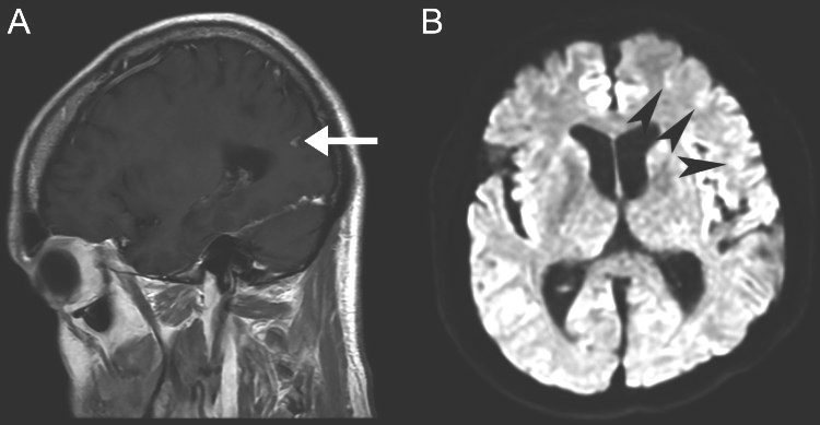
Brain MRI - sagittal T1-weighted post-contrast (A) and axial diffusion-weighted (B) images
Tiny enhancing foci in the left parietal lobe cortex (arrow) and tiny acute to subacute cortical infarcts at the left frontal lobe were favored to represent enhancing tiny subacute infarcts
MRI: magnetic resonance imaging