Brain Sciences 2024 (Nov 7); 14 (11): 1124
Figure 8.
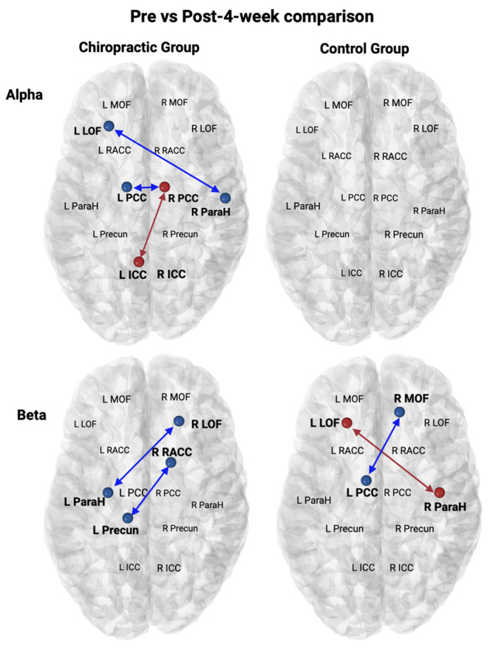
Pre- vs. post 4 weeks comparisons within both groups in SEPs. A significant increase in connectivity is shown as red and a decrease in connectivity is shown as blue. R ICC—Right Isthmus Cingulate Cortex; L ICC—Left Isthmus Cingulate Cortex; R LOF—Right Lateral Orbitofrontal; L LOF—Left Lateral Orbitofrontal; R MOF—Right Medial Orbitofrontal; L MOF—Left Medial Orbitofrontal; R PCC—Right Posterior Cingulate Cortex; L PCC—Left Posterior Cingulate Cortex; R Precun—Right Precuneus; L Precun—Left Precuneus; R ParaH—Right Parahippocampal cortex; L ParaH—Left Parahippocampal Cortex; R RACC—Right Rostral Anterior Cingulate Cortex; L RACC—Left Rostral Anterior Cingulate Cortex.