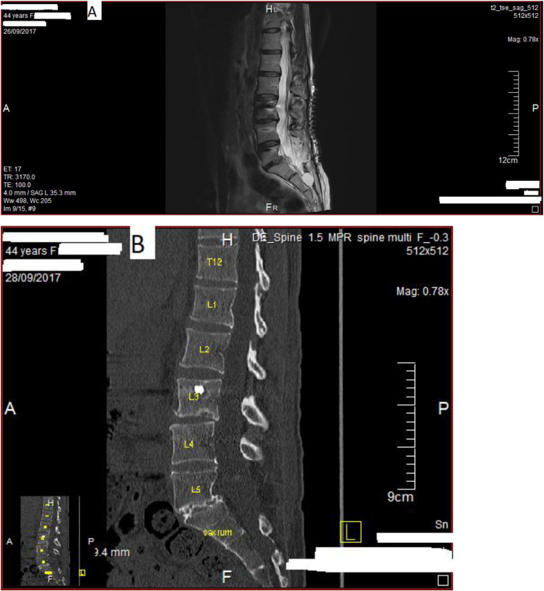
Baseline MRI findings of the case. (A) Baseline sagittal T2-weighted lumbosacral magenetic resonance (MR) image of the case after lumbar stabilization revision operation at L3-L5 level on September 2017, depicting bulging at L3-L4 level. In addition, there is defect in posterior elements, including fat infiltration in multifidus at L2-S1 level. Heterogenous and edematous paraspinal muscles and loss of lumbar lordosis are seen at L3-S1 level. Metalic sutures on the skin are also seen. Note that the distinct structures in the vertebral bodies of L3-L5 are bilateral internal transpedicular screw rod system. At the sacral area, a Tarlov cyst is seen. (MR date: September 26, 2017.) (B) Baseline sagittal lumbosacral computed tomographic (CT) image of the case after lumbar stabilization revision operation at L3-L5 level in September 2017, depicting degenerative changes and osteophytes at L3-L4 and especially at L5-S1 level. Moreover, there is grade I spondylolisthesis at L5-S1 level and loss of lumbar lordosis. Note that white heart-shaped area at the vertebral body of L3 is a part of internal transpedicular screw rod system. (CT date: September 28, 2017.)