PMC full text:
Published online 2022 Oct 20.
Figure 3
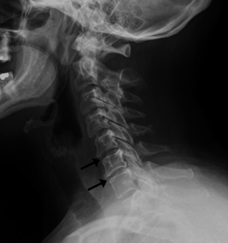
Cervical spine radiograph, lateral view, from 10 years prior to presentation.
The radiograph shows reduction in disc spacing at C5-6 and C6-7 with marginal anterior osteophyte formation consistent with spondylosis (arrows). In other views (not shown), uncovertebral joint hypertrophy was evident on the right at C5-6 and on the left at C6-7 producing a mild degree of foraminal encroachment bilaterally.