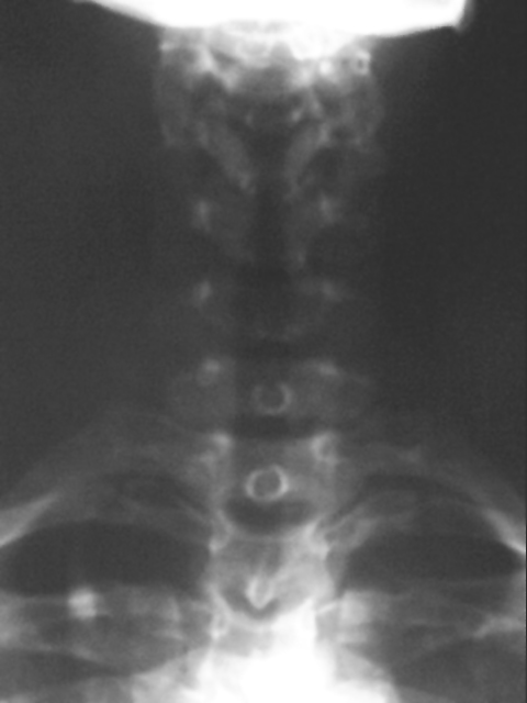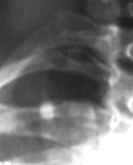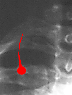

The Azygos Lobe


By G. Patrick Thomas, Jr., DC, DACBR
This normal variant occurs in less than 5% of the population. Despite it's characteristic shadow, it often alarms physicians not familiar with it's apearance. The azygos lobe forms when the azygos vein fails to migrate over the apex of the lung during fetal life. Instead, it courses through the lung, dragging along with it the parietal and visceral pleura. The four layers of pleura are then known as the 'azygos fissure', and the bit of lung tissue separated from the rest of the lung is known as the 'azygos lobe'. The azygos vein itself is seen on end, and may simulate a pulmonary mass. This normal variant is often seen on AP lower cervical views, as was the situation with this particular case. It's important to note that this anomoly is of no clinical significance-just don't confuse it with other more serious conditions.
Click the images below for a close-up:


Return to RADIOLOGY


| Home Page | Visit Our Sponsors | Become a Sponsor |
Please read our DISCLAIMER |