This report describes the development of specialized pattern analysis software that accepts temperature data exported from the TyTron Thermographic instrument. The TyTron is a dual probe infrared thermographic instrument that provides temperature information for the skin on the left and right sides of the spine. The user can compare scans from the left side, the right side, and the delta or side-to-side difference. The newly developed software provides tools for manipulation and visualization of two overlapping plots and for calculation of congruence between thermographs taken on different occasions.
Using the pattern analysis software, the user can shift and crop the thermographs to produce the best subjective overlap before calculating congruence factors. After the scans are prepared for analysis, five congruence factors are calculated using a variety of algorithms. All of the calculations are scaled to produce factors from 0 to 1, where 1 is perfect congruence. The calculated factors are area ratio, slope comparison, correlation coefficient, and slope and amplitude comparisons using methods similar to those of Stewart and Boone. The pattern analysis software is being used in studies of thermographic pattern stability at Sherman College and Life University.
KEYWORDS: Chiropractic, Skin Temperature, Thermography, Pattern Analysis
Chiropractors and medical thermographers have developed differing methods for interpreting paraspinal thermograms. Detailed scans of whole regions, as provided by liquid crystal or video thermography can be used to locate unusually warm or cool regions. The approach to interpretation in this case is to compare like locations along the back, looking for symmetry of temperature. Here, asymmetry is considered abnormal in proportion to the magnitude of the temperature difference. Wallace, Wallace, and Resh surveyed the extensive literature on paraspinal thermography and described the thermoregulative physiology behind its use as a clinical assessment tool (9).
Chiropractors often use paraspinal thermograms from dual probe devices. These devices provide temperature information only on thin strips of skin on either side of the spine. Commonly, these devices only provide information on the difference between side-to-side temperatures without plotting the actual skin temperature. One method of analysis is to locate the presence of "breaks," where there is a sharp side-to-side swing of the temperature trace. DeBoer, et al. (10) and Plaugher, et al. (11) tested the inter- and intra-examiner reliability of differential paraspinal temperature recording and analysis. Plaugher used a thermocouple device that made contact with the skin and tested the ability of operators to detect the same breaks. DeBoer, using a noncontact infrared thermal device, was able to digitize the thermograms and compare temperature data statistically. In both studies, the authors found agreements ranging from fair to substantial by using the Intraclass Correlation Coefficient as a basis for comparison.
Other methods have been devised for analyzing differential thermograms, including the counting of "constants" or similar regions of the graph that reappear over time (12). In this type of pattern analysis, the contour or character of the temperature plot is considered more important than the magnitude of the temperature swing from the centerline. Recently, Hart and Boone developed a method for assessing the degree of similarity between thermograms using a manual measurement system where the proportion by length of regions of the temperature pattern that had similar slopes was used to calculate a percentage of pattern (4). In this method, the degree of similarity of thermograms is based largely on the analyzer's judgement, although some measurement or counting is also involved.
In 1989, Stewart, Riffle, and Boone noted the need for an objective assessment of temperature pattern, especially to produce reliable measures for research (13). They developed mathematical methods for analysis of digitized thermograms, although, at the time, no such digitized temperature patterns were available to them. In the intervening years, digital thermographic systems that provide numeric output of skin temperature profiles have become widely available. Owens, et al. developed computational methods that make use of such digitized thermograms (14). Computer-aided pattern analysis is useful and convenient because it quickly provides a numeric assessment of the degree of similarity of thermograms and allows clinicians to detect patterns in an objective fashion. This report describes the pattern analysis software developed at Sherman College of Straight Chiropractic.
Paraspinal thermographs for this project were recorded using the TyTron C-3000 Infrared thermal scanner (Titronics Research & Development, Oxford, Iowa) interfaced to an MS-Windows-based computer. The TyTron is a hand-held dual probe scanner with wheels to keep it a uniform distance from the skin during data collection. One of the wheels is equipped with a position sensor that tracks its location along the spine as the temperature is recorded. For scans, the patient is seated on a special backless chair. The full spine thermal scan is a continuous glide from the second sacral tubercle to the shelf of the occiput. A typical full-spine scan can be collected in 15 seconds. Thermal data are stored in a patient database for later viewing and interpretation. For the purposes of this study, Titronics Research & Development provided us with a special export routine from their software that saves the centigrade temperature data in a comma delimited text file.
Software tool development
Our goal in developing software to help interpret thermal scans was to provide an objective and efficient method for comparing any two scans. The temperature file consists of two columns of temperature data, one for each side of the spine. The number of records depends on the length of the patient's spine and varies generally between 300 and 500 data pairs. (The TyTron collects data at a rate of 6 points per centimeter.) Temperature comparison, therefore, could involve comparing the left temperature profiles on two occasions, the right profiles, or a calculated left/right difference, called the delta profile.
We developed the software tools for this project in Microsoft Visual BASIC 6.0 (VB6), which combines good graphing capabilities and mathematical command libraries. Our approach was to provide general graphing and calculation tools, but to let the operator control the flow of the program. The initial steps of preconditioning, in particular, involve the operator's judgement and visual interpretation. The intent was to use the operator to produce the best visual overlap of data files before congruence is calculated.
Preconditioning: Normalization
Previous studies showed that the temperature of the back changes with time, depending on how long the patient is allowed to equilibrate to room temperature. In one study, the back temperature was seen to change continuously for 30 minutes, although certain features of the scans became stable after 9 minutes (15). For analysis purposes, we desired to remove artifacts from the data that might have to do with the amount of time the patient was allowed to equilibrate with differences in ambient temperature. We developed a preconditioning algorithm, normalizing the temperature file on the basis of the range of temperatures found in the data file, to remove variability due to absolute temperature differences between scans. Essentially, before the comparison of similarity occurs, the data points are all shifted so that the highest and lowest temperatures are normalized between the two files. Hence, a new data set is calculated from the temperature file wherein the range of temperatures between highest and lowest covers the same span in both data sets. The left probe, right probe, and delta (difference) information are each normalized separately. Normalized data files are graphed in a vertical orientation from the cervical to the sacral spine. Color-coding is used to distinguish data files from each other. The two data files that are being compared are referred to as file1 and file2.
Sliding and clipping
Early observation of the thermal scan data indicated that TyTron operators did not always begin scans at precisely the same level of the spine or end them at the same spot. Hence, temperature scans performed on the same patient are not usually precisely the same length, and the locations of characteristic peaks do not always occur at the same record in the data set. Two more preconditioning stages were developed to handle artifacts having to do with starting and ending regions of the scans. A clipping tool can be used to crop off areas of the traces at the beginning and end if necessary. The operator decides where to crop the files. Cropped areas are not considered in normalization or congruence calculations. A sliding tool was also developed to enable the operator to move one trace up or down with respect to the other in an attempt to visually produce the best overlap of the traces. After graphing the normalized plots, the operator can observe certain characteristic peaks where the traces overlap well. If the peaks clearly do not line up, the operator uses the sliding tool to move one trace up in increments of 1 step with respect to the other. Because the TyTron has a built-in position sensor, the number of data points in successive scans varies little and seems to vary only when the starting and stopping points are different. Hence, we saw no need to scale or normalize the data sets with respect to length.
Smoothing
In some cases the temperature plot shows erratic changes. The delta plot in particular, because it is the difference between two changing profiles, is prone to this effect. We added a smoothing tool to the software to make comparisons between plots easier when the information is erratic. The smoothing tool calculates a new data set with a moving average algorithm that sets the value of each data point equal to the average of the 4 data points on each side of it in the data file. Certain of the congruence calculations described below are sensitive to the slope of the temperature plot and produce calculation errors when the temperature plot is perfectly vertical. Smoothing is necessary in those cases to allow the calculation to proceed through that segment.
Congruence Calculations
The software operator uses the congruence calculation tools after he or she is satisfied that all the preconditioning steps have produced a pair of traces that are most likely to provide the best similarity. Several different analytical methods are used to detect patterns in thermal scans by chiropractors. Similarly, a wide variety of mathematical methods might be used. We decided to provide a sampling of different methods, some of which compare the temperature scans on a global basis, and some of which compare small adjacent sections to each other. All of the calculated congruence factors are scaled so that perfect congruence provides a factor of 1.00.
Area Ratio
The simplest mathematical calculation of congruence involves simply taking the area between the two curves. The first step is to sum the absolute value of the difference between the normalized temperature values (between file1 and file2) at each vertical location in the trace. In the second step, the area ¾ bounded by a rectangle as tall as the length of the files, and as wide as their range ¾ is calculated. The area ratio is the area of the difference between the files subtracted from the total area of the bounding rectangle and divided by the total area. Perfectly congruent curves will have no area between them and produce an area ratio of 1.00; the area ratio will decrease from there, as curves become more disparate.
Slope Comparison
The slope comparison algorithm calculates the instantaneous slope at each point along the temperature profiles and compares the slope of one file to the other at that point. The instantaneous slope for point i along the data file is calculated as:
where NTemp is the normalized temperature.
The slope of the ith point of File1 is compared with the slope of the ith point of File2. If the slopes are within a threshold value (Slope Sensitivity) set by the operator, then the two temperature files are considered congruent at that point. The comparison is done for every point on the temperature plot, and the number of congruent points is tallied. The slope comparison reported to the operator is the ratio of the number of congruent points to the number of points in the data file. Hence, a slope comparison on identical files produces a value of 1.00. The slope comparison can be made as stringent as desired by varying the Slope Sensitivity value. The default value of 1.00 makes a very sensitive test for congruence.
Correlation
The correlation coefficient calculation is another global comparison of temperature profile congruence. The equation used is that for Pearson's product moment, r:
where X and Y are the normalized temperature values for File1 and File2 respectively and n is the number of points in the data file.
A scattergram plot of File1 versus File2, displayed as the calculation is performed, shows how well the two files compare. Very similar temperature scans show a tight grouping of (x,y) pairs along a diagonal line.
Stewart/Boone methods
Stewart, Riffle, and Boone developed two statistical methods for comparing temperature scans (9). They used a piecewise comparison wherein segments 10-data-points long were compared on the basis of amplitude and slope. This statistical method uses tests to determine the probability of two segments being from the same population, and has a confidence interval of 95%. We used two similar methods in developing our software and dubbed them Stewart/Boone Methods r% and t%. In our adaptation of these methods, we also used 10-data-point-long segments of the temperature data files. The algorithm steps through the data file and compares overlapping 10-data-point segments. The ith segment will compare data points i-4 through i+5. Hence, each segment overlaps with the next adjacent segment by 9 points.
In the slope comparison (r%), the Pearson product moment (r) is calculated by comparing 10-data-point segments of File1 with 10-data-point segments of File2. The value of r is evaluated for significance by comparison to a statistical table. (In this case, the degrees of freedom (df) = 8 and r > 0.632 are significant at the 0.05 level.) All the overlapping segments are tested for congruence in turn and the proportion of the data file that is congruent is presented as a fraction, where 1.00 represents perfect congruence. In the amplitude comparison (t%), the Student's t-test is used to compare 10-data-point segments of File1 with the same segment of File2. The calculated value of 't' is compared with values in a table to determine significance. (For df = 8, t>1.734 is significant at the 0.05 level.). Again, the proportion of the data files that are congruent is presented as a number from 0 to 1.00.
Interpretation of congruence calculations
Because this method of analysis is new, we do not yet know how to interpret the congruence values. Already we can see that some factors are more sensitive than others: the same input files produce a range of congruence factors. For instance, the area ratio tends to produce high values whereas the slope comparison is more sensitive.
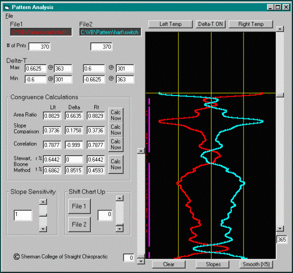
One way to test the sensitivity of the calculations is to use specially prepared test files. In one set of experiments, actual temperature scan files were reversed, i.e., the left and right probe data were swapped. Switching the input order in this way produces delta plots that are the reciprocals of each other. Fig 1 shows a screen capture of the pattern software with one of the switched files plotted and analyzed. The resulting congruence factors for a set of 7 runs on switched data files produced an area ratio on these files, which are about as different from each other as can be accomplished, averaging 0.53 (Table 1). The Correlation coefficient shows a value of negative 1, as expected. The Stewart/Boone methods show an interesting result. The calculation based on slope similarity (r%) produced 0 for all 7 files tested, whereas the calculation based on amplitude was less sensitive, producing a range of values between 0.41 and 0.69 (avg 0.53).
Fig 1. A screen capture of the pattern software being used to calculate congruence factors for a switched data file. The right and left probe data have been swapped in the data set, producing a delta channel in which the File1 and File2 data sets are reciprocal. This method was used to determine the relative sensitivity of the different congruence factors.

Table 1. Summary table of calculated congruence factors for 7 files where the left and right probe data are switched, producing reciprocal delta values.
| Area Ratio | Slope | Correlation | r % | t % | |
| Avg | 0.532 | 0.148 | -1.000 | 0.000 | 0.526 |
| Max | 0.773 | 0.202 | -0.999 | 0.000 | 0.691 |
| Min | 0.361 | 0.006 | -1.000 | 0.000 | 0.407 |
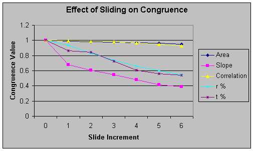
A second test of the sensitivity was performed by testing the effects of sliding on the congruence factors. In this experiment, congruence factors were calculated when each of 3 actual data files was compared to itself, i.e. the same file was loaded into File1 and File2. In the experiment, File1 was translated six steps up with respect to File2 in increments of one step at a time. The congruence factors were calculated at each step.
Fig 2. The effect of sliding on congruence. Identical files were tested for congruence, then slid in increments of one step with respect to the other. The TyTron records approximately six data points per centimeter of travel so that six steps in the data set would represent one centimeter of displacement of the temperature profile. The slope comparison shows the greatest sensitivity to sliding, while area ratio and correlation vary very little.

Table 2. The effects of sliding on the congruence factors. Identical files were tested for congruence, then slid in increments of one step with respect to the other. This table shows the average values for three different data file sets. The slope comparison shows the greatest sensitivity to sliding, and area ratio and correlation vary the least.
| Slide | Area | Slope | Correlation | r % | t % |
| 0 | 1.000 | 1.000 | 1.000 | 1.000 | 1.000 |
| 1 | 0.987 | 0.678 | 0.998 | 0.936 | 0.864 |
| 2 | 0.982 | 0.605 | 0.992 | 0.831 | 0.841 |
| 3 | 0.975 | 0.550 | 0.982 | 0.741 | 0.725 |
| 4 | 0.967 | 0.482 | 0.968 | 0.662 | 0.607 |
| 5 | 0.960 | 0.419 | 0.952 | 0.600 | 0.558 |
| 6 | 0.952 | 0.386 | 0.933 | 0.547 | 0.542 |
The area ratio and correlation factors were relatively insensitive to sliding (Table 2 and Figure 2), decreasing from 1 to 0.93 when one file was slid six steps with respect to the other. Slope comparison and the Stewart/Boone methods were more sensitive to sliding, decreasing from 1 to 0.54 (0.38 for slope comparison) with six steps of sliding. Six steps in the data set represents 1 centimeter of travel of the scanning gun along the patient's back.
In clinical use, this software would be used to determine the degree of similarity of temperature profiles to help determine when an adjustment is needed. We still need to determine which congruence factor works best in the clinical setting and what range of values tell us when "pattern" is present. Reliability studies of the software are currently being carried out at Sherman College and Life University. Serial thermographic readings will show how much change occurs in the temperature patterns over the short term and what changes can be expected with adjustment. The calculated congruence factors will lend an element of objectivity to the results of those studies.
Another possible use of the system and a way to "calibrate" the congruence factors would be to have a panel of experts compare the calculations subjectively to determine if a pattern exists. Thermograms can be printed on paper and distributed to a group of experienced chiropractors for analysis. The chiropractors can use their own preferred method to indicate which scans show the presence of pattern, and perhaps to what degree pattern is present. With that information, it should be possible to calibrate the congruence factors, or provide cut-off values, so that novice chiropractors can use the objective numbers generated as an indication of the presence of pattern in their patient's thermograms.