

Clinical Management of a Veteran with Chronic
Scan-negative Cauda Equina Syndrome:
A Case ReportThis section is compiled by Frank M. Painter, D.C.
Send all comments or additions to: Frankp@chiro.org


FROM: J Canadian Chiropractic Assoc 2026 (Aug); 70 (2): 236–248 ~ FULL TEXT
OPEN ACCESS Zachary E. Scott, DC • Kevin W. Meyer, DC • Clinton J. Daniels, DC, MS
White City VA Medical Center,
University of Western States,
White City, Oregon, USA.
Objective: To describe the clinical presentation, diagnostic considerations, and safety-first course of chiropractic care for an adult male with chronic scan-negative cauda equina syndrome.
Clinical features: A 41-year-old male U.S. Veteran sought chiropractic care for progressively intensifying chronic low back pain and bilateral lower extremity pain along with progressive bladder dysfunction, groin hypoesthesia, and sexual dysfunction. The history and examination findings raised concern for cauda equina syndrome, but a same-day MRI and neurosurgery consult failed to yield surgical indications.
Intervention and outcomes: Chiropractic care involved 8 visits over 10 weeks. Care focused on neurological monitoring and patient education along with soft-tissue therapy, flexion-distraction, and spinal manipulation. The patient reported improvements in pain, function, and neurological features following this course of care, and no adverse events occurred.
Summary: This case emphasizes timely recognition of features associated with cauda equina syndrome and illustrates a cautious, multimodal approach to chiropractic co-management for an individual with scan-negative cauda equina syndrome.
Keywords: Veterans health; cauda equina syndrome; chiropractic; low back; manual therapies; pain; veterans health services.
From the FULL TEXT Article:
Background
Seronegative spondyloarthropathies are a group of chronic, immune-mediated inflammatory diseases that affect between 0.5-1.9% of individuals worldwide, and which are responsible for considerable pain, disability, social burden, and decreased quality of life. [1] Axial spondyloarthritis (axSpA) is the most common seronegative spondyloarthropathy subtype and is widely underdiagnosed. An estimated 300,000 Canadians live with axSpA and nearly two-thirds of them experience significant symptoms. [2]
Classically, axSpA has a familial predilection, is highly associated with the human leukocyte antigen B27 (HLA-B27) allele, and is also most commonly diagnosed in males younger than 45 years old. [3] Clinical symptoms include chronic inflammatory back pain (IBP) and stiffness, peripheral arthralgia, enthesitis, and extra-articular complications such as psoriasis, as well as symptoms of the ocular, gastrointestinal, and pulmonary systems. [4]
While this condition was formerly described exclusively as ankylosing spondylitis, differentiation between subtypes of this condition was prompted by the increased availability of magnetic resonance imaging (MRI) which paved the way for earlier detection of sacroiliitis and axSpA recognition before other visible radiographic changes (e.g., ankylosis). The enhanced sensitivity in detection evolved the understanding of the disease course and prompted a 2022 update in the Assessment of Spondyloarthritis International Society (ASAS) classification and nomenclature to create a distinction between the most common subtypes of this condition, radiographic axial spondyloarthritis (r-axSpA) and non-radiographic axial spondyloarthritis (nr-axSpA). [5–7]
Management recommendations for patients with axSpA include a personalized approach with a multidisciplinary team coordinated by a rheumatologist. In addition to medication management with non-steroidal anti-inflammatory drugs (NSAIDs) and biologic medications, non-pharmacological approaches like therapeutic exercise, patient education, and lifestyle change are commonly recommended. [7, 8] Chiropractic services (e.g., advice, education, spinal manipulation, manual therapies, exercise prescription) are well documented for their benefits to patients with chronic mechanical low back pain and are recommended by clinical practice guidelines for the management of back pain. [9, 10] Patients with axSpA do present to chiropractors seeking treatment, [11] however, literature pertaining to chiropractic management of axSpA remains limited. [12–15]
In the US and Canada, chiropractic is a doctoral-level profession that holistically approaches the evaluation and management of musculoskeletal conditions with non-surgical and non-pharmacological approaches. [16] The purpose of this case series is to describe the chiropractic management of three patients at different progressions of axSpA disease diagnosis and management within an integrated hospital-based system.
Case presentations
We followed the CARE Checklist guidelines for reporting for case reports. [17]
Case 1
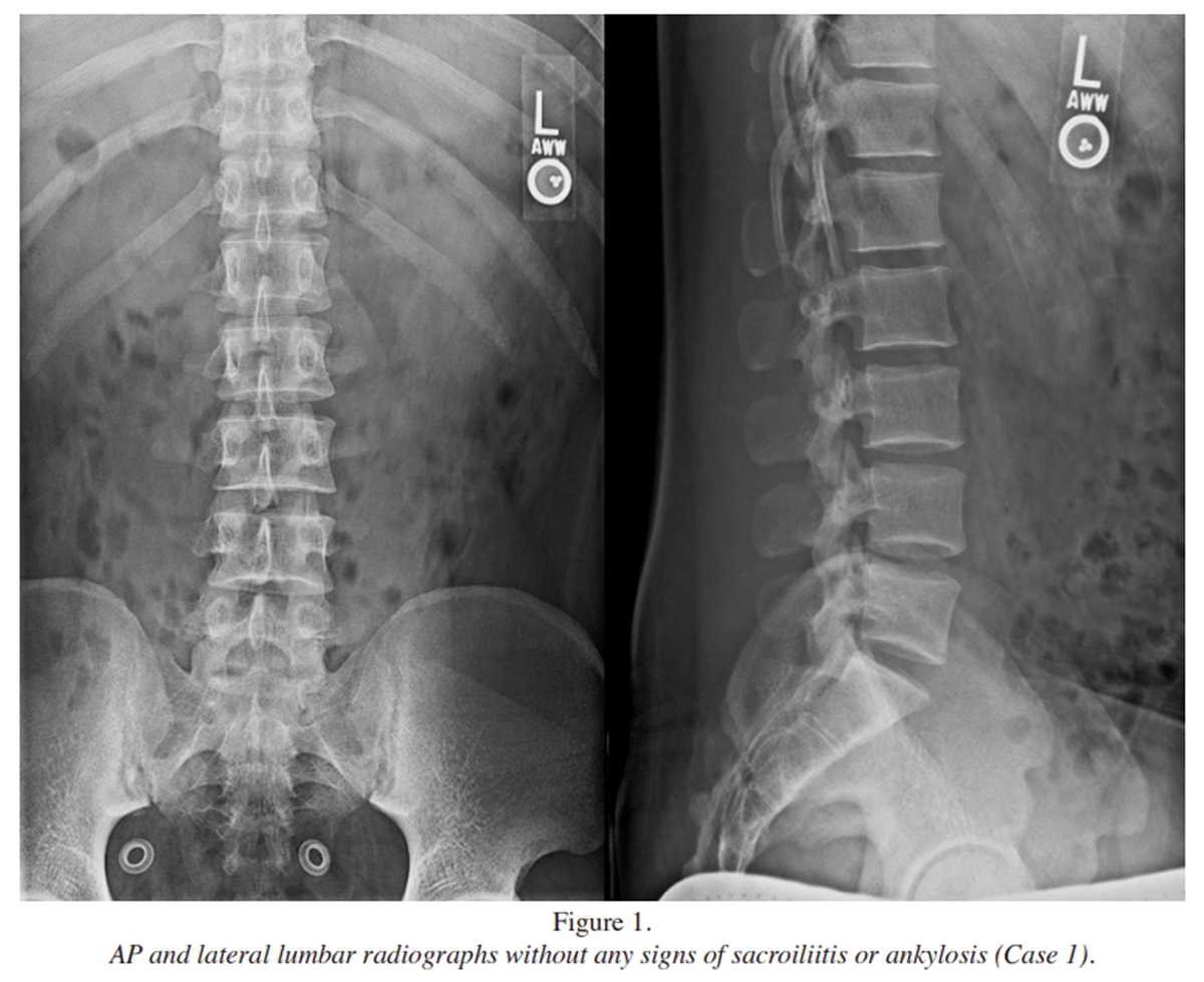
Figure 1 A 22-year-old Hispanic male U.S. Army veteran presented to the chiropractic clinic with a history of chronic lower back and left posterolateral hip pain. Two years earlier, he was diagnosed with nr-axSpA and was medically discharged from military service. A chart review of a rheumatology consultation one year before the chiropractic consultation revealed chronic uveitis, laboratory testing demonstrating the presence of HLA-B27 allele and elevated inflammatory markers, and advanced imaging with MRI confirmed bilateral sacroiliitis. Ankylosis was not visible on plain-film lumbar radiographs. (Figure 1)
He did not have known family history of autoimmune disease, toxic exposure, or additional pertinent past medical history. Relevant treatment history included minimal benefit with oral NSAIDs and steroid injections to the SI joints, and significant relief of pain and stiffness after initiating rheumatologist-prescribed the Tumor Necrosis Factor inhibitor (TNFi) medication adalimumab.
Upon presentation to the chiropractic clinic, he described symptoms of diffuse joint stiffness upon waking and following heavy lifting as well as pain and stiffness which went through cycles of relapsing and remitting. Pain intensity was rated 2 out of 10, while interference with enjoyment of life and general activity were rated 2 and 1 out of 10, respectively using the pain, enjoyment of life, and general activity (PEG) scale. [18] He reported daily morning stiffness, lasting for up to three hours, that was relieved by walking and stretching. His referral was prompted by a six-month relapse of lower back and left hip pain that hindered his ability to lift weights, run, and play soccer.
Upon examination, pathological reflexes were absent and muscle stretch reflexes, lower extremity myotomes, and skin sensation were all within normal limits. Lumbar extension was moderately reduced and provocative for pain at the bilateral posterior superior iliac spine while lumbar flexion was palliative. Prone SI joint extension bilaterally provoked ipsilateral lower back pain. Thigh thrust and lumbar facet loading were unremarkable. Pain and tenderness were elicited upon palpation of the left quadratus lumborum, left hip external rotators, and the bilateral SI joints. The patient was diagnosed in the chiropractic clinic with mechanical nonspecific lumbosacral pain, complicated by nr-axSpA. As no red flags were observed, the veteran was actively following rheumatology, and a recent pelvic MRI and laboratory testing were on file, no additional workup was deemed appropriate at the time of consultation.
Given the absence of ankylosis, a trial of care was initiated with thoracolumbar, lumbopelvic, and bilateral femoroacetabular high-velocity low-amplitude (HVLA) manipulation, flexion-distraction manipulation to the lumbar spine, myofascial release to the lumbar and hip musculature, and a targeted home exercise program promoting lumbopelvic stability for eight visits over six months.
The patient was educated regarding the relapsing-remitting nature of systemic inflammatory conditions. He was encouraged to stay active, including a graded approach to returning to weightlifting, running, and playing soccer, and he was reassured that increasing his activity level to tolerance was safe without undue risk of setting back his overall improvement. He was also advised to continue with treatment as recommended by the rheumatologist to minimize symptoms and disease progression.
As expected, his symptoms did relapse and remit throughout the course of care. During these relapses, the veteran reported difficulty connecting with his prescribing provider to refill his biologic medication. The treating chiropractor contacted the patient’s rheumatologist to facilitate refill of the prescription.
Following a therapeutic withdrawal from care, a re-evaluation was performed at visit 8. At this time, the veteran presented with minimal lower back pain, with intensity rated 1 out of 10 (50% improvement), and pain impact on enjoyment of life and general activity were rated 0 (100% improvement) and 1 (no change) out of 10, respectively. Although pain flares in the absence of medication continued, he resumed weightlifting three to four times weekly with improved tolerance, regularly playing soccer, and began practicing yoga.
He denied experiencing any soreness or adverse events related to his chiropractic treatments. At this time, he was transitioned to care on an as-needed basis for flare management, and it was recommended that he continue his home exercise program.
Case 2
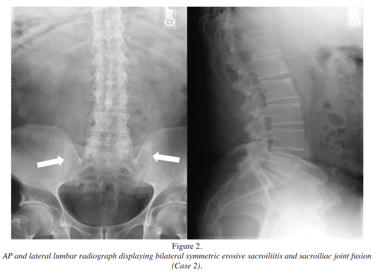
Figure 2 A 55-year-old white male U.S. Navy veteran with previously diagnosed r-axSpA presented to the chiropractic clinic with chronic non-progressive neck, mid back, and lower back pain with intermittent radiating pain to the right groin and posterior knee. He reported his symptoms began after contracting salmonellosis thirty-five years earlier, which led to frequent attacks of diffuse axial and peripheral joint pain since the infection. Nine years later, he tested positive for HLA-B27, was diagnosed with chronic irritable bowel disease, and lumbar radiographic imaging revealed bilateral symmetric erosive sacroiliitis and fusion of the SI joints. (Figure 2)
No ankylosis was evident within the lumbar spine. Other pertinent radiographic imaging revealed mild multilevel degeneration of the cervical spine, mild anterior wedging of T8, T9, and T11 within the thoracic spine, and femoral neck osteopenia with a dual-energy X-ray absorptiometry (DEXA) scan performed two years earlier.
He did not have any known history of toxic exposure or other pertinent past medical history. His recent treatment regimen included rheumatologist-prescribed TNFi (infliximab) infusions every five weeks for the last seven years. He described infusions as crucial to reducing his frequency and intensity of symptomatic exacerbations and additionally perceived that his chiropractic treatments played an important role in his ability to perform activities of daily living and reduced his need for oxycodone medication which was prescribed to be used on an as needed basis.
Due to patient reported benefit with chiropractic treatment, for the past twelve years he was managed with a supportive care plan consisting of patient education, manual therapy, exercise, and lifestyle advice for chronic pain.
At his most recent trial of care, pain intensity, enjoyment of life, and general activity were all rated 5 out of 10 using the PEG scale, and Neck Disability Index [19] was scored 6 out of 40 (two questions were skipped by the patient). He denied any temporal association with pain in all regions of complaint and reported thoracic pain that was provoked with deep inspiration and coughing. Upon examination, pathological reflexes were absent, and muscle stretch reflexes, upper extremity myotomes, and skin sensation were all within normal limits. Cervical, thoracic, and lumbar ranges of motion were all moderately reduced, except for left thoracic rotation, which was markedly decreased, and all ranges of motion increased his pain and stiffness.
Facet loading and prone SI extension testing both increased lumbosacral pain bilaterally. Repeated end-range loading with cervical retraction and lumbar extension were palliative for local pain. The veteran’s goals for treatment were to obtain manual therapies and self-management strategies for continued pain relief and to minimize the need for pain medication. He was diagnosed with mechanical nonspecific cervical, thoracic, and lumbosacral pain, complicated by multiple thoracic compression deformities, r-axSpA, and reactive arthritis. As the veteran was actively following rheumatology and his symptoms were stable at time of presentation, obtaining updated imaging and labs to evaluate inflammatory activity were deemed unnecessary.
Treatment consisted of cervical, cervicothoracic, thoracic, and lumbar HVLA spinal manipulation as well as myofascial release to each region of complaint. In addition to his medication management prescribed by rheumatology, he was prescribed and encouraged to pursue a consistent home exercise program of postural-based strengthening and stretching, and to begin walking for exercise on a consistent basis.
Throughout this most recent trial of care, significant improvement in thoracic and lumbosacral mobility was observed without adverse event reported. At re-evaluation pain was rated 7 out of 10 (40% worsening), enjoyment of life was rated 5 out of 10 (no change), and general activity was rated 6 out of 10 (20% worsening), using the PEG scale, reflective of the veteran experiencing a flare-up of lumbopelvic pain.
As recommended during treatment, he initiated a consistent home exercise program, reporting he had lost 10 pounds over 5 months. At his request, we agreed to a supportive chronic pain treatment plan at a frequency of one visit every one to two months.
Case 3
A 36-year-old white male U.S. Army veteran was referred to the chiropractic clinic for chronic neck and lower back pain, and he additionally reported poor sleep quality and depression. Prior to the chiropractic presentation, rheumatology was consulted due to suspicion of axSpA. Chart review revealed that the patient was positive for the HLA-B27 allele and suffered from chronic uveitis and peripheral arthralgias. The patient denied a family history of autoimmune disease, symptoms of inflammatory bowel disease, or psoriatic skin lesions.
An MRI performed six years earlier was unremarkable for evidence of SI joint inflammation or ankylosis. Considering the absence of MRI findings, the rheumatologist assessed that there was a low suspicion of seronegative spondyloarthropathy and diagnosed him with fibromyalgia. Two years later, following worsening chronic uveitis with unclear etiology, the patient returned to rheumatology. He was prescribed a short dose of the biologic medication adalimumab, and he experienced significant relief of his ocular symptoms and musculoskeletal pain but was lost to follow-up.
He presented to the chiropractic clinic one year later, reporting atraumatic diffuse neck pain and worsening, insidious, lower back pain accompanied by morning stiffness lasting for three to four hours on average. His pain alternated in laterality at the lumbosacral junction and bilateral posterolateral hips. He also reported pain radiating to the right posterior thigh, without paresthesia or weakness. Pain intensity was rated 7 out of 10, while impact on enjoyment of life was rated 6 out of 10, and generalized activity was rated 8 out of 10. Neck Disability Index was scored 32 out of 50, reflecting severe disability.
He reported using a single-point cane when walking long distances due to lower back pain and stiffness. Prior treatment with lumbar radiofrequency ablation provided him with relief, but symptoms were now regressed back to pre-treatment status.
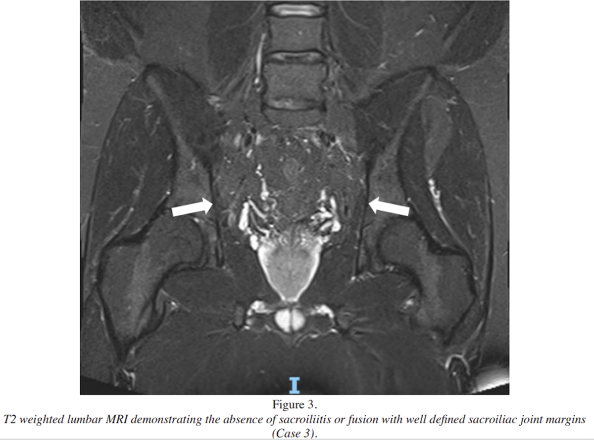
Figure 3 Upon examination, pathological reflexes were absent, and muscle stretch reflexes, upper and lower extremity myotomes, and skin sensation were all within normal limits. Cervical, thoracic, and lumbar ranges of motion were all moderately reduced and provocative for pain. Passive supine cervical rotation was provocative for neck pain bilaterally, while no other cervical orthopedic testing elicited symptoms. Lumbar facet loading increased ipsilateral lumbosacral pain. Posterior pelvic pain provocation increased SI joint pain ipsilaterally to the side that was tested. He was diagnosed with mechanical nonspecific cervical, thoracic, and lumbar pain with concern for axSpA. Updated lumbar and pelvic MRIs were ordered, and coordination efforts were initiated to reconnect the veteran with rheumatology to inquire about re-establishing treatment with adalimumab. MRI revealed a mild symmetric L5-S1 disc bulge without nerve root contact, and signs of sacroiliitis remained absent. (Figure 3)
The chiropractic treatment plan consisted of cervical, cervicothoracic, thoracic, and lumbosacral HVLA spinal manipulation, myofascial release, and a home exercise program with an emphasis on cervical and thoracic mobilization, as well as pelvic stabilization exercises. He was followed in the clinic once per month for 18 months. Throughout this care, the veteran presented with fluctuating pain intensity, which became more stable after re-establishing care with a new rheumatologist. That provider diagnosed the veteran with recurrent uveitis with concern for inflammatory peripheral arthritis and/or nr-axSpA and re-initiated adalimumab medication.
At final re-evaluation, the patient noted reduced stiffness in all regions of complaint following re-initiation of the biologic medication and a decrease in pain intensity and improved ability to perform generalized activity, which were rated 5 (29.5% improvement) and 7 (13% improvement) out of 10 respectively on the PEG questionnaire. At this point, he was transitioned to an as-needed treatment plan for flare-up management. He denied experiencing any soreness or adverse events related to his chiropractic management.
Discussion
This case series presents the first description of chiropractic management for US military veterans with known or suspected axSpA spanning various stages of disease progression.
Case 1 represented the management of an early progression of nr-axSpA in a younger veteran who received multidisciplinary care including prescription medication from rheumatology, and manual therapy, exercise, and education from the chiropractor.
Case 2 described supportive care for chronic pain in an older veteran with longstanding r-axSpA who received consistent chiropractic care for chronic pain, complementing his primary rheumatologic treatment.

Table 1
page 8
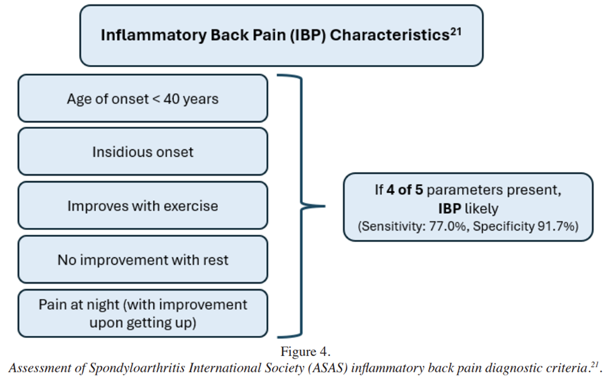
Figure 4
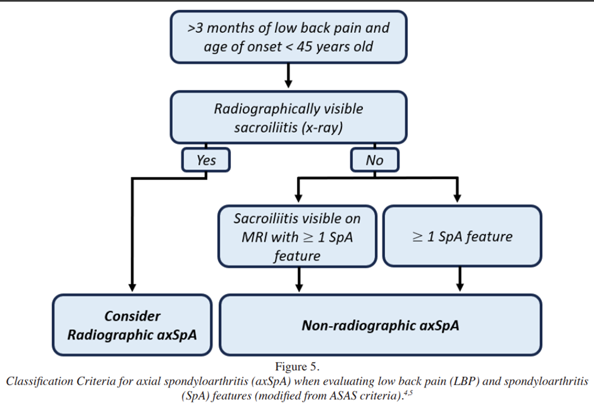
Figure 5 Case 3 involved diagnostic testing and coordination of care for a patient with suspected axSpA. In the absence of other disease states, recognition of well-established clinical features greatly increases the likelihood of identifying axSpA. [20] (Table 1)
Chronic IBP is a key clinical feature in differentiating axSpA from other conditions. Hallmarks of IBP include relapsing and remitting back pain of insidious onset that begins before age forty, is worse at night and upon first waking, improves with movement, and does not improve with rest (Figures 4 and 5). [21, 22] Although the specificity of IBP for an axSpA diagnosis is limited, if these hallmark symptoms are present, then further investigation is indicated and referral to a rheumatologist is warranted. [23]
All three cases exhibited IBP symptoms, tested positive for HLA-B27, and presented extraspinal features, but disease manifestation visible on imaging varied. Despite radiographic features being absent in patients with nr-axSpA, evidence of inflammatory lesions such as bone marrow edema and fat metaplasia within the SI joints may be present and viewed with MRI. Rather than two distinct diseases, the non-radiographic distinction in nomenclature functions to capture the broad spectrum of axSpA and allow for acknowledgment of disease progression. [24]
A considerable number of cases with nr-axSpA may never develop radiographic sacroiliitis or ankylosis. [25] When transition from nr-axSpA to r-axSpA does occur, it tends to progress slowly, occurring in only 5.8% of patients over 10 years, with even lower rates in those managed with TNFis. [25] The ASAS recommends rheumatologist opinion for axSpA diagnosis, and their classification criteria combine clinical, laboratory, and imaging features.
The differential diagnoses for axSpA share many similar features and can be difficult to distinguish between. Alternative diagnostic considerations include mechanical non-specific low back pain, spondylosis, fibromyalgia, diffuse idiopathic skeletal hyperostosis (DISH), Scheuermann disease, osteitis condensans ilii, and infectious sacroiliitis. [21] Diagnostic imaging evidence of inflammatory sacroiliitis on MRI is a suggestive of axSpA, but other conditions can mimic this appearance. Conditions presenting similarly on imaging include osteoarthritis, stress fractures, infective sacroiliitis, and osteitis condensans ilii. [26]
Radiographic diagnosis can be further complicated as sacroiliitis is not specific for axSpA as it has been observed in patients with mechanical low back pain and must be correlated with other clinical findings alongside expert opinion by rheumatologist to confirm diagnosis. [27] The HLA-B27 allele is highly associated with axSpA, but has also been found to be prevalent in those with standalone anterior uveitis and inflammatory bowel disease, juvenile idiopathic arthritis, reactive arthritis, psoriatic arthritis, and undifferentiated spondyloarthritis. [28] Many of these diagnoses may be present concurrently and they are not mutually exclusive.
Case 3 was initially diagnosed with fibromyalgia, only to receive medication treatment relief two years later as nr-axSpA was considered. [29] In the absence of definitive axSpA imaging findings or lab markers such as HLA-B27, c-reactive protein, and erythrocyte sedimentation rate, differentiating between the two pathologies can be difficult. The prevalence of fibromyalgia has been reported to co-occur with axSpA in between 4-25% of cases. [30]
Case 2 presented with a past medical history of reactive arthritis, a disease in the same family of seronegative spondyloarthropathies as axSpA. Inflammatory sacroiliitis is predictive for reactive arthritis disease progression, with 22-26% of cases evolving to r-axSpA. [31, 32] Disc degeneration is also common in patients with axSpA and may provoke concurrent mechanical spine pain. [33] Case presentations may have similar clinical characteristics on clinical examination, laboratory findings, or diagnostic imaging, but they often do not fit the comprehensive clinical profile of axSpA.
According to the ASAS guideline, the entirety of the clinical picture must be accounted for alongside expert opinion by rheumatologist to confirm diagnosis. [7] The development of ankylosis of the spine is the leading source of functional limitation in patients with established axSpA. [34] Despite the wide variation of severity of inflammatory symptoms and degree of ankylosis, the disease burden and management are similar for both nr-axSpA and r-axSpA. [35]
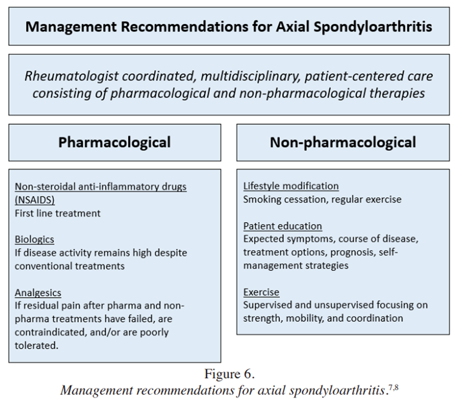
Figure 6 The goals of treatment are generally to alleviate symptoms, improve functioning, maintain the ability to work, decrease disease complications, and forestall skeletal damage as much as possible. Clinical guidelines recommend treatment with medications such as NSAIDs, TNFis, supervised and unsupervised exercise, and formal education on self-management and the nature of the disease. [7, 8] (Figure 6)
Chiropractors employ various methods to address axial pain, including joint manipulation and mobilization, soft-tissue therapies, patient education, lifestyle recommendations, therapeutic exercise, and physical modalities (e.g., electrical stimulation, heat). Although disease presentations varied and the diagnosis of one case was uncertain, chiropractic management of each case remained similar, was guideline-concordant, and, as an adjunct to medication management, may have contributed to reduced pain symptoms, improved function, attainment of functional goals, and increased enjoyment of life.
Chiropractic services do not address the underlying inflammatory etiology of axSpA, but they are consistent with management guidelines and may play a role in pain management, improving mobility, and improving function. A recently published observational study suggests that more education of chiropractors is needed to increase appropriate screening for inflammatory back pain in their offices. [11] In cases where IBP is suspected, chiropractors can play a role in referral coordination to a rheumatologist, as was done in cases one and three.
The ability of chiropractors to function in the larger healthcare system as licensed independent practitioners can support patients’ pain and quality of life through recognition of axSpA and providing guidance towards appropriate management indirectly or directly alongside a rheumatologist. Chiropractic providers must be cautious of potential safety concerns when treating patients with axSpA, particularly when applying spinal manipulation. [36, 37]
Chronic inflammation, limited spinal mobility, and increased physical inactivity with axSpA can lead to both new bone formation in the spine as well as increased bone loss. This bone loss represents a point of caution when providing HVLA spinal manipulation, as it is well established that patients with axSpA have a high prevalence of osteoporosis and fracture alongside potential ankylosis. [38–41]
The American College of Rheumatology (ACR) recommends against spinal manipulation for patients with r-axSpA in the presence of spinal fusion or advanced spinal osteoporosis. [8] No recommendations were made by ACR in the context of spinal manipulation and nr-axSpA. In contrast, the World Health Organization guidelines on safety in chiropractic do not consider ankylosing spondylitis to be contraindicated in the absence of ligamentous instability, anatomic subluxation, or ankylosis. [10] Our case 2 with r-axSpA did have known osteopenia via a recent DEXA scan. However, the severity of osteopenia was mild, and he had a long history of benefit with spinal manipulation.
Therefore, treatment using HVLA spinal manipulation was initiated, and no adverse events occurred. Fracture risk should be considered by the treating chiropractor and clinical decisions regarding spinal manipulation are best considered on a case-by-case approach.
Limitations
Our report has several limitations inherent to its design as a case series. Case series do not have a control group which precludes the ability to establish causality between treatment administered and the observed outcomes described. As these patients were treated in an outpatient setting, several uncontrolled factors exist that may have influenced their response to treatment.
Treatments did not occur in isolation, and it is not possible to identify which treatment(s) resulted in patient improvements. Further, low back pain episodes are commonly self-limiting and axSpA is relapsing and remitting in nature, thus natural history may have played a role in the outcomes reported. [42, 43]
We suspect that appropriate medication management of IBP symptoms led to the largest effect on patient outcomes. The role of the chiropractors may be most well suited to identifying IBP, coordination with rheumatology, and providing adjunctive non-pharmacologic treatment options. This study was also limited to a small sample size that may not be generalizable to other patients with axSpA.
Summary
This case series suggests that chiropractic services may be effective as an adjunct to standard medical care in patients with spine pain due to Axial spondyloarthritis (axSpA). While it is important for providers to be cautious of potential contraindications to HVLA spinal manipulation, such as osteoporosis and spinal fusion from ankylosis, none were present in these patients and chiropractic care was well-tolerated without adverse events. Further research is needed to investigate the role of chiropractors in the management of individuals with chronic spine pain related to axSpA.
Abbreviations
ASAS = Assessment of Spondyloarthritis International Society
axSpA = axial spondyloarthritis
DEXA = dual energy X-ray absorptiometry
DISH = diffuse idiopathic skeletal hyperostosis
HLA-B27 = human leukocyte antigen B27
HVLA = high-velocity, low-amplitude
IBP = inflammatory back pain
MRI = magnetic resonance imaging
nr-axSpA = non-radiographic axial spondyloarthritis
NSAID = non-steroidal anti-inflammatory drug
PEG = pain, enjoyment of life, and general activity
r-axSpA = radiographic axial spondyloarthritis
SI = sacroiliac joints
SpA = spondyloarthritis
TNFi = tumor necrosis factor inhibitor
Authors’ contributions
ZS, KM, and CD drafted the manuscript which was critically revised and approved by all authors and provided patient care described within this study.
References:
Bakland G, Nossent HC.
Epidemiology of Spondyloarthritis: A Review.
Curr Rheumatol Rep. 2013;15(9):351.
doi:10.1007/s11926-013-0351-1Ankylosing Spondylitis – Symptoms, Causes, Diagnosis & Treatments.
Accessed November 13, 2025. https://arthritissocietycanada.org/about-
arthritis/arthritis-types-(a-z)/types/ankylosing-spondylitisFeldtkeller E, Khan M, Van Der Heijde D, Van Der Linden S, Braun J.
Age at disease onset and diagnosis delay in HLA-B27 negative vs. positive patients with ankylosing spondylitis.
Rheumatol Int. 2003;23(2):61-66.
doi:10.1007/s00296-002-0237-4Schwartzman S, Ruderman EM.
A Road Map of the Axial Spondyloarthritis Continuum.
Mayo Clin Proc. 2022;97(1):134-145.
doi:10.1016/j.mayocp.2021.08.007Rudwaleit M, Landewe R, Van Der Heijde D, et al.
The development of Assessment of SpondyloArthritis international Society classification criteria for axial spondyloarthritis (part I): classification of paper patients by expert opinion including uncertainty appraisal.
Ann Rheum Dis. 2009;68(6):770-776.
doi:10.1136/ ard.2009.108217Rudwaleit M, Van Der Heijde D, Landewe R, et al.
The development of Assessment of SpondyloArthritis international Society classification criteria for axial spondyloarthritis (part II): validation and final selection.
Ann Rheum Dis. 2009;68(6):777-783.
doi:10.1136/ard.2009.108233Ramiro S, Nikiphorou E, Sepriano A, et al.
ASASEULAR recommendations for the management of axial spondyloarthritis: 2022 update.
Ann Rheum Dis. 2023;82(1):19-34.
doi:10.1136/ard-2022-223296Ward MM, Deodhar A, Gensler LS, et al.
2019 Update of the American College of Rheumatology/Spondylitis Association of America/Spondyloarthritis Research and Treatment Network Recommendations for the Treatment of Ankylosing Spondylitis and Nonradiographic Axial Spondyloarthritis.
Arthritis Care Res. 2019;71(10):1285-1299.
doi:10.1002/acr.24025Rubinstein SM, de Zoete A, van Middelkoop M, Assendelft WJJ, de Boer MR, van Tulder MW.
Benefits and Harms of Spinal Manipulative Therapy for the
Treatment of Chronic Low Back Pain: Systematic Review
and Meta-analysis of Randomised Controlled Trials
British Medical Journal 2019 (Mar 13); 364: 1689World Health Organization.
WHO Guideline for NonSurgical Management of Chronic Primary
Low Back Pain in Adults in Primary and Community Care Settings
World Health Organization; 2023.Deodhar A, Bhalerao S, Kiwalkar S, et al.
Prevalence of axial spondyloarthritis (axSpA) in patients treated for chronic back pain in chiropractic clinics: the oregon chiropractic axial spondyloarthritis study (ORCAS).
Clin Rheumatol. 2025;44(10):3981-3991.
doi:10.1007/s10067- 025-07655-xDeodhar A, Kiwalkar S, Mehendale T, Bhalerao S.
Axial Spondyloarthritis in the Chiropractic Care Setting: A Systematic Literature Review.
JCR J Clin Rheumatol. 2022;28(2):e589-e595.
doi:10.1097/ RHU.0000000000001776Cornelson SM, Beavers D, Harvey A, Hogarth W, Kettner NW.
Chiropractic Care in the Management of Inactive Ankylosing Spondylitis: A Case Series.
J Chiropr Med. 2017;16(4):300-307.
doi:10.1016/j.jcm.2017.10.002Rose KA, Kim WS.
The effect of chiropractic care for a 30-year-old male with advanced ankylosing spondylitis: a time series case report.
J Manipulative Physiol Ther. 2003;26(8):524-532.
doi:10.1016/S0161-4754(03)00110-6Rutherford SM, Nicolson CF, Crowther ER.
Symptomatic improvement in function and disease activity in a patient with ankylosing spondylitis utilizing a course of chiropractic therapy: a prospective case study.
J of the Canadian Chiropractic Association. 2005 Jun;49(2):81-91.
PMID: 17549197; PMCID: PMC1840019.LeFebvre R, Peterson D, Haas M.
Evidence-Based Practice and Chiropractic Care
J Evid Based Comp Altern Med. 2012 (Dec 28); 18 (1): 75–79Riley DS, Barber MS, Kienle GS, et al.
CARE guidelines for case reports: explanation and elaboration document.
J Clin Epidemiol. 2017;89:218-235.
doi:10.1016/j.jclinepi.2017.04.026Krebs EE, Lorenz KA, Bair MJ, et al.
Development and Initial Validation of the PEG, a Three-item Scale Assessing Pain Intensity and Interference.
J Gen Intern Med. 2009;24(6):733-738.
doi:10.1007/s11606-009-0981-1Vernon H, Mior S.
The Neck Disability Index:
A Study of Reliability and Validity
J Manipulative Physiol Ther 1991 (Sep); 14 (7): 409–415Van Gaalen FA, Rudwaleit M.
Challenges in the diagnosis of axial spondyloarthritis.
Best Pract Res Clin Rheumatol. 2023;37(3):101871.
doi:10.1016/j.berh.2023.101871Walsh JA, Magrey M.
Clinical Manifestations and Diagnosis of Axial Spondyloarthritis.
JCR J Clin Rheumatol. 2021;27(8):e547-e560.
doi:10.1097/RHU.0000000000001575Sieper J, van der Heijde D, Landewé R, et al.
New criteria for inflammatory back pain in patients with chronic back pain: a real patient exercise by experts from the Assessment of SpondyloArthritis international Society (ASAS).
Ann Rheum Dis. 2009;68(6):784-788.
doi:10.1136/ard.2008.101501Dubreuil M, Sieper J.
Inflammatory back pain and axial spondyloarthritis: lessons for clinical practice and epidemiological research.
Arthritis Rheumatol Hoboken NJ. 2018;70(7):981-983.
doi:10.1002/art.40462Michelena X, López-Medina C, Marzo-Ortega H.
Nonradiographic versus radiographic axSpA: what’s in a name?
Rheumatol Oxf Engl. 2020;59(Suppl 4):iv18-iv24.
doi:10.1093/rheumatology/keaa422Molto A, López-Medina C, Sepriano A, et al.
Sacroiliac radiographic progression over 10 years in axSpA: data from the DESIR inception cohort.
Ann Rheum Dis. 2024;83(7):858-864.
doi:10.1136/ard-2023-225184Tsoi C, Griffith JF, Lee RKL, Wong PCH, Tam LS.
Imaging of sacroiliitis: Current status, limitations and pitfalls.
Quant Imaging Med Surg. 2019;9(2):318-335.
doi:10.21037/qims.2018.11.10Arnbak B, Grethe Jurik A, Hørslev-Petersen K, et al.
Associations Between Spondyloarthritis Features and Magnetic Resonance Imaging Findings: A Cross-Sectional Analysis of 1,020 Patients With Persistent Low Back Pain.
Arthritis Rheumatol. 2016;68(4):892-900.
doi:10.1002/art.39551Gill T, Rosenbaum JT.
Putative Pathobionts in HLAB27-Associated Spondyloarthropathy.
Front Immunol. 2021;11:586494.
doi:10.3389/fimmu.2020.586494Derry S, Wiffen PJ, Häuser W, et al.
Oral nonsteroidal anti-inflammatory drugs for fibromyalgia in adults.
Cochrane Database Syst Rev. 2017;2017(3):CD012332.
doi:10.1002/14651858.CD012332.pub2Mease PJ.
Fibromyalgia, a missed comorbidity in spondyloarthritis: prevalence and impact on assessment and treatment.
Curr Opin Rheumatol. 2017;29(4):304-310.
doi:10.1097/BOR.0000000000000388BANICIOIU-COVEI S, VREJU FA, CIUREA P.
Predictive Factors for the Evolution of Reactive Arthritis to Ankylosing Spondylitis.
Curr Health Sci J. 2015;41(2):104-108.
doi:10.12865/CHSJ.41.02.03Inman RD, Chiu B.
Early cytokine profiles in the joint define pathogen clearance and severity of arthritis in Chlamydia-induced arthritis in rats.
Arthritis Rheum. 2006;54(2):499-507.
doi:10.1002/art.21643Bittar M, Deodhar A.
Axial Spondyloarthritis: A Review.
JAMA. 2025;333(5):408-420.
doi:10.1001/jama.2024.20917Landewe R, Dougados M, Mielants H, Van Der Tempel H, Van Der Heijde D.
Physical function in ankylosing spondylitis is independently determined by both disease activity and radiographic damage of the spine.
Ann Rheum Dis. 2009;68(6):863-867.
doi:10.1136/ard.2008.091793López-Medina C, Ramiro S, van der Heijde D, Sieper J.
Characteristics and burden of disease in patients with radiographic and non-radiographic axial Spondyloarthritis: a comparison by systematic literature review and meta-analysis.
RMD Open. 2019;5(2):e001108.
doi:10.1136/rmdopen-2019-001108Whalen WM, Hawk C, Farabaugh RJ, et al.
Best Practices for Chiropractic Management of Adult
Patients With Mechanical Low Back Pain: A Clinical
Practice Guideline for Chiropractors
in the United States
J Manipulative Physiol Ther 2022 (Oct); 45 (8): 551-565Gatterman M.
Standards of practice relative to complications of and contraindications to spinal manipulative therapy.
J Can Chiropr Assoc. 1991;35(4):232-236.Lim MJ, Kang KY.
A Contemporary View of the Diagnosis of Osteoporosis in Patients With Axial Spondyloarthritis.
Front Med. 2020;7:569449.
doi:10.3389/fmed.2020.569449Nigil Haroon N, Szabo E, Raboud JM, et al.
Alterations of bone mineral density, bone microarchitecture and strength in patients with ankylosing spondylitis: a cross-sectional study using high-resolution peripheral quantitative computerized tomography and finite element analysis.
Arthritis Res Ther. 2015;17:377.
doi:10.1186/s13075-015-0873-1Macêdo MB, Shinjo SK, Domiciano DS.
Breaking the diagnosis: ankylosing spondylitis evidenced by cervical fracture following spine manipulation.
Intern Emerg Med. 2022 Jan;17(1):273-274.
doi: 10.1007/s11739-021-02829-2. Epub 2021 Aug 20.
PMID: 34415490.Liao CC, Chen LR. Anterior and Posterior Fixation of a Cervical Fracture Induced by Chiropractic Spinal Manipulation in Ankylosing Spondylitis: A Case Report.
J Trauma Inj Infect Crit Care. 2007;63(4):E90-E94.
doi:10.1097/01.ta.0000246957.22573.63Fritz JM, Rhon DI, Teyhen DS, et al.
A Sequential Multiple-Assignment Randomized Trial (SMART) for Stepped Care Management of Low Back Pain in the Military Health System: A Trial Protocol.
Pain Med Malden Mass. 2020;21(Suppl 2):S73-S82.
doi:10.1093/pm/pnaa338Bodunrin A.
What is the Natural History of Low Back Pain? It Depends – Duke Center of Excellence in Manual and Manipulative Therapy.
March 14, 2025. Accessed November 13, 2025.
https://sites.duke.edu/cemmt/2025/03/14/wha
Return to LOW BACK PAIN
Return to CHIROPRACTIC CARE FOR VETERANS
Since 10-04-2026
| Home Page | Visit Our Sponsors | Become a Sponsor |
Please read our DISCLAIMER |